| Table of Contents |  |
|
Clinical Image
| ||||||
| Primary non-Hodgkin's lymphoma of liver | ||||||
| Daniel Vasilev Kostov1, Georgi Leonidov Kobakov2, Daniel Veselov Yankov3 | ||||||
|
1MD, PhD, MSc Professor of Surgery, Director, Department of Surgery, Naval Hospital of Varna, BG-9010 Varna, 3 Hristo Smirnenski Street, Bulgaria.
2MD, PhD, Professor of Surgery, Director, Department of Oncologic Surgery, Division of Surgery, Marko Markov Interregional Dispensary and Hospital of Oncological Diseases, BG-9010 Varna, 3 Hristo Smirnenski Street, Bulgaria. 3MD, PhD, Department of Surgery, Naval Hospital of Varna, BG-9010 Varna, 3 Hristo Smirnenski Street, Bulgaria. | ||||||
| ||||||
|
[HTML Abstract]
[PDF Full Text]
[Print This Article]
[Similar article in Pumed] [Similar article in Google Scholar] |


| How to cite this article: |
| Kostov DV, Kobakov GL, Yankov DV. Primary non-Hodgkin's lymphoma of liver. Int J Hepatobiliary Pancreat Dis 2014;4:48–51. |
|
Case Report
| ||||||
|
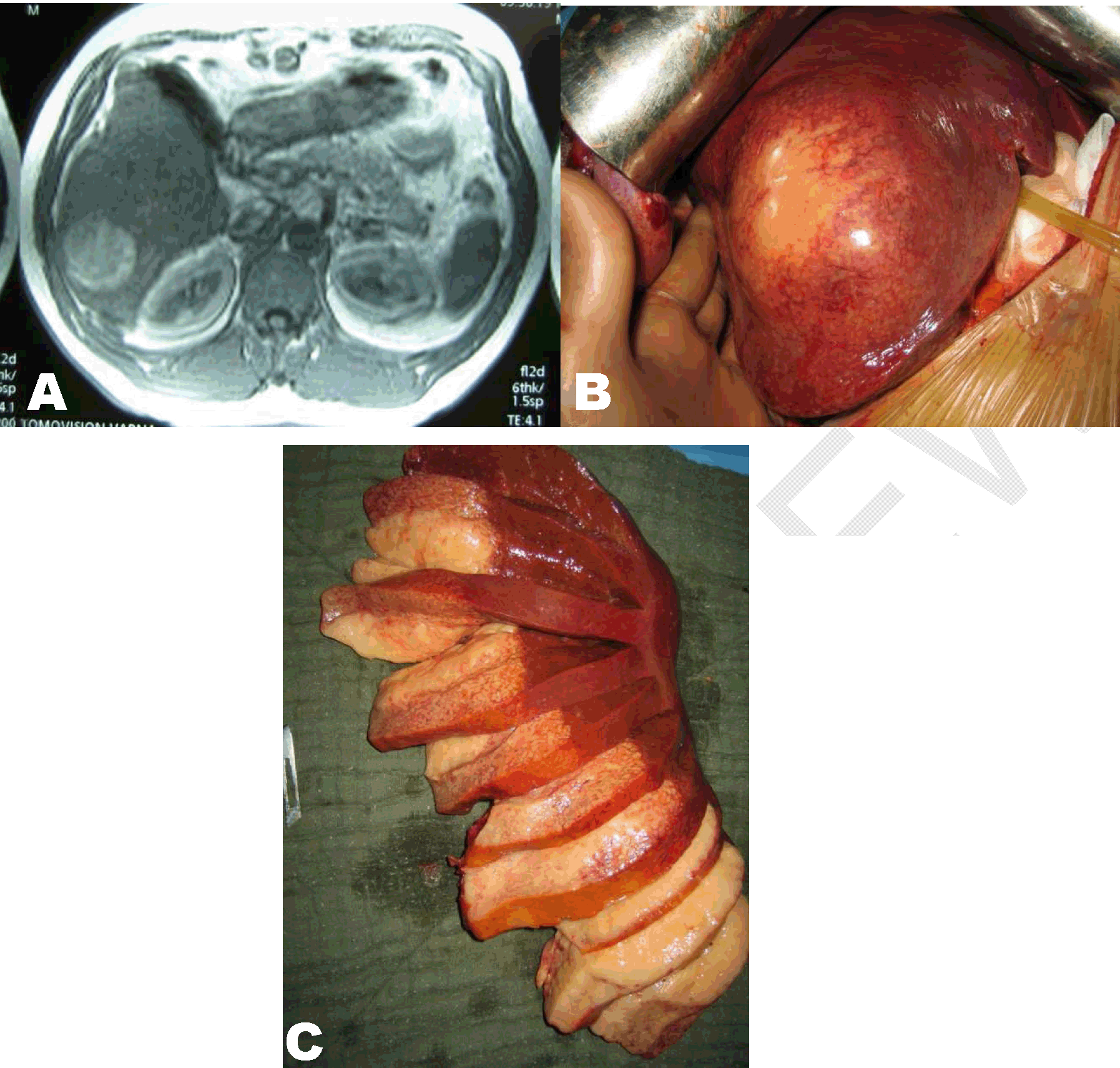
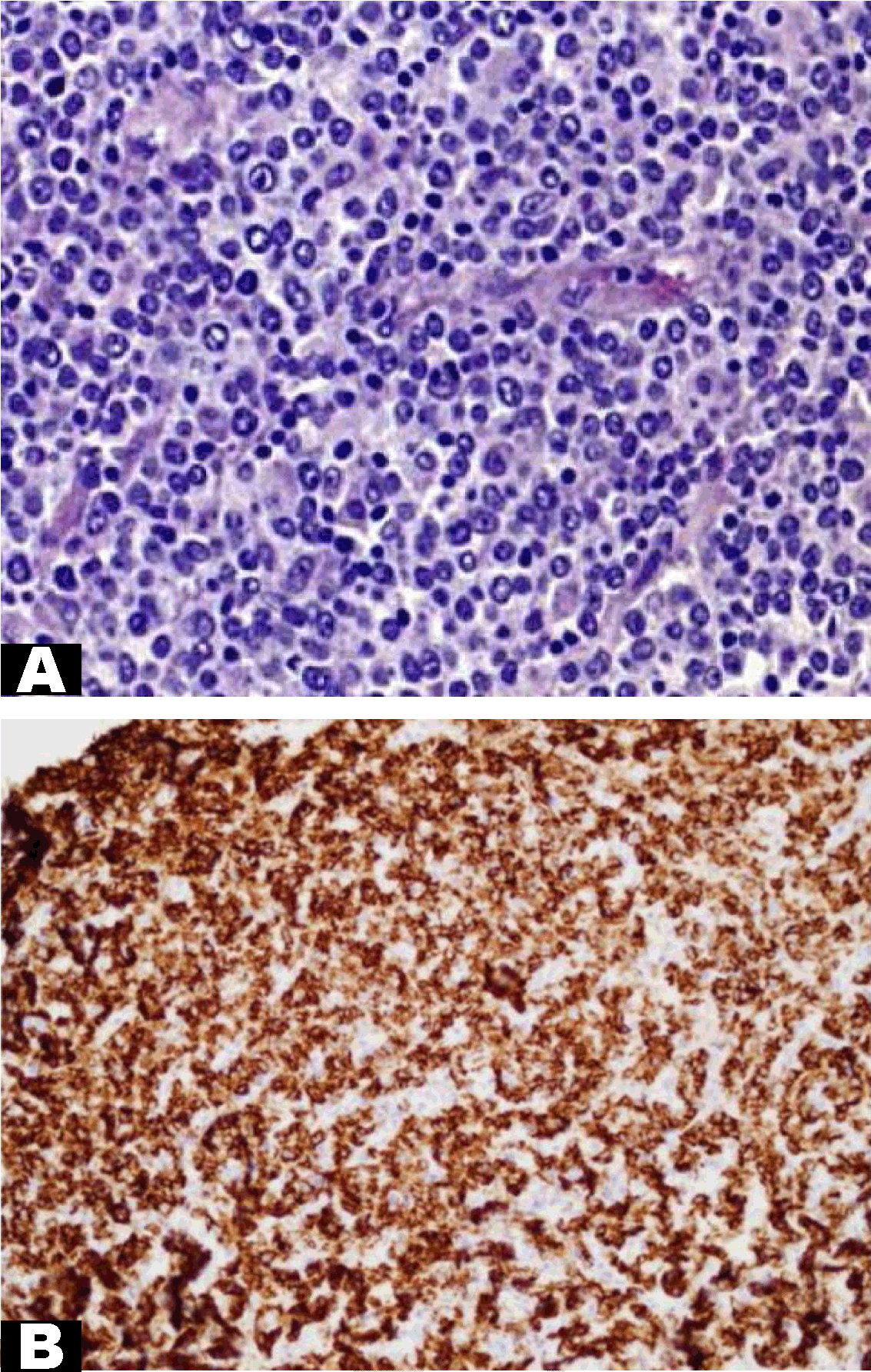
A 65-year-old male was presented his primary care physician with a two-month history of constipation, upper right abdominal pain and occasional right groin pain. His past medical history included hypertension and hepatitis B from which he had recovered (HBsAg negative). The biochemical analyses revealed alanine transaminase 35 U/L, aspartate transaminase 38 U/L and bilirubin level 26 mmol/L with normal blood count. The result of quantitative HBV DNA revealed an undetectable viral load which is generally lower than 300 copies/mL. Magnetic resonance imaging (MRI) scan of the abdomen showed a mass in the right lobe of the liver with a diameter of 10 cm, located on the right hemiliver (Figure 1A). The tumors markers CA19–9 was 57.3 IU/mL (normal range <27 IU/mL), carcinoma antigen 15–3 was 29.6 IU/mL (normal range <5 IU/mL), and CA125 was 72.4 IU/mL (normal range <35 IU/mL). Alpha-fetoprotein and carcinoembryonic antigen were within normal range. Liver biopsy confirmed non-Hodgkin's lymphoma, staining positive for CD20 lymphocytes. The patient was diagnosed with primary large B cell lymphoma of the liver stage IE (stage I), given that no additional foci of lymphoma were found anywhere else in the body. A decision was made that the patient had to undergo an operation. Intraoperative findings showed a large tumor mass with a diameter of 10 cm in the right hemiliver which necessitated a right hemihepatectomy (Figure 1B-C). A histomorphological examination of the resected specimen showed large lymphoid cells varying in shape from oval to round vesicular nuclei containing fine chromatin (Figure 2A). The immunohistochemical findings were positive for the pan-B cell marker CD20, as well as LCA, and CD43. Immunostaining of the liver lesion showed reactivity for CD3, CD5, CD10, CD138, MUM1, bcl6, and bcl2 (Figure 2B). Ki67 (MIB-1) immunostaining detected a high proliferation fraction of lymphoid cells. A highly malignant B cell non-Hodgkin's lymphoma diagnosis was given. A liver resection was followed by four cycles of chemotherapy with cyclophosphamide 750 mg/m2, doxorubicin 50 mg/m2, vincristine 1.4 mg/m2, prednisone 100 mg/day, rituximab 375 mg/m2 (CHOP-R). One year postoperatively, the patient is in good clinical condition without a recurrence of the lymphoma. | ||||||
|
| ||||||
|
| ||||||
| ||||||
|
Discussion
| ||||||
|
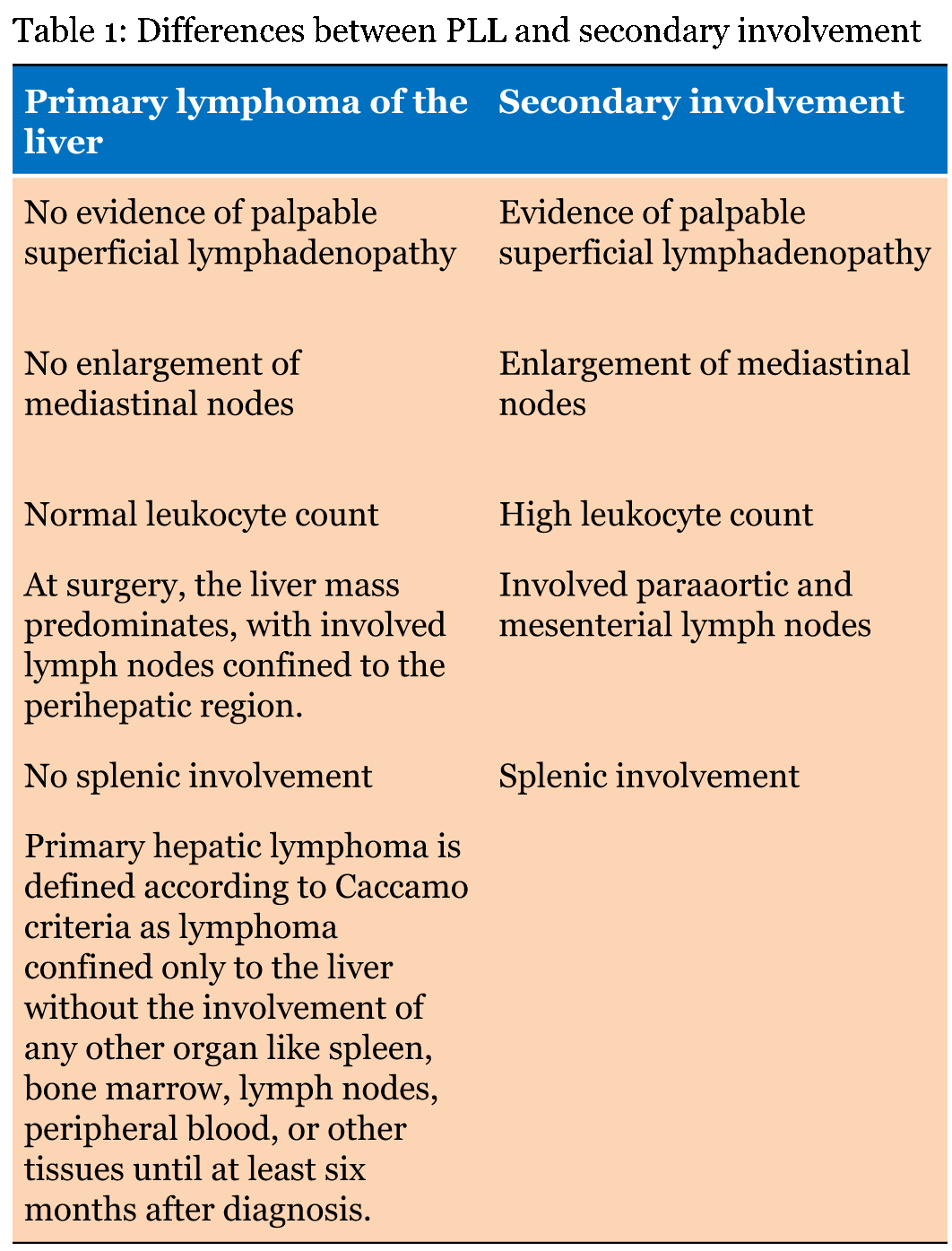
Primary hepatic lymphoma (PHL) constitutes about 0.4% of the cases of extra nodal non-Hodgkin's lymphoma, and only about 0.016% of all cases of non-Hodgkin's lymphoma [1]. While the pathogenesis of PHL is currently unclear there is evidence that it has been associated with Epstein-Barr virus, hepatitis C virus, human immunodeficiency virus, or human T-lymphotropic virus infections, liver cirrhosis, systemic lupus erythematosus, and immunosuppressive therapy [2]. The tumor arises from lymphoid elements in the liver, and it is defined by the following clinical criteria (Table 1):
Extranodal lymphoma is classified as secondary if there is involvement of lymph nodes, except for those of an adjacent primary organ or with more than one extra nodal site. The diffused large B cell is the most common type of non-Hodgkin's lymphoma, and more than 50% of patients have some extra-nodal lesions [3]. To diagnose a PHL it is necessary for the liver biopsy to coincident with lymphoma and an absence of a lymphoproliferative disease outside the liver. Late diagnosis, however, or failure to make a diagnosis can end up in fulminant hepatic failure and ultimately death. The optimal treatment for PHL has not yet been defined. Many reports have suggested that surgical resection followed by adjuvant chemotherapy and/or radiation results in better prognosis [4]. The current indications that necessitate a surgery are cases of patients with a localized disease that can undergo radical liver resection or if there is presence of a persistent respectable disease followed by chemotherapy [5]. Chemotherapy with CHOP-R based regimens is the gold standard [6]. | ||||||
| ||||||
|
| ||||||
|
Conclusion
| ||||||
|
Primary non-Hodgkin's lymphoma of the liver is a rare variation of lymphoma and making a precise diagnosis presents a challenge in clinical practice. Magnetic resonance imaging findings of primary and secondary lymphoma may not be sufficient to render a specific diagnosis, especially when liver involvement is the first or only finding. Rather, other parameters must be considered, including a liver biopsy with a histopathological examination. | ||||||
|
References
| ||||||
| ||||||

|
[HTML Abstract]
[PDF Full Text]
|
|
Author Contributions
Daniel Vasilev Kostov – Substantial contributions to conception and design, Acquisition of data, Analysis and interpretation of data, Drafting the article, Revising it critically for important intellectual content, Final approval of the version to be published Georgi Leonidov Kobakov – Analysis and interpretation of data, Revising it critically for important intellectual content, Final approval of the version to be published Daniel Veselov Yankov – Analysis and interpretation of data, Revising it critically for important intellectual content, Final approval of the version to be published |
|
Guarantor of submission
The corresponding author is the guarantor of submission. |
|
Source of support
None |
|
Conflict of interest
Authors declare no conflict of interest. |
|
Copyright
© 2014 Daniel Vasilev Kostov et al. This article is distributed under the terms of Creative Commons Attribution License which permits unrestricted use, distribution and reproduction in any medium provided the original author(s) and original publisher are properly credited. Please see the copyright policy on the journal website for more information. |
|
|
|
About The Authors
| |||
| |||
| |||
| |||