| Table of Contents |  |
|
Review Article
| ||||||
| Focal nodular hyperplasia and hepatocellular adenomas: What is new in 2013? | ||||||
| Elisa Palladino1, Daniele Sommacale2, Renaud Siboni1, Christine Hoeffel3, Christian Lechner1, Tullio Piardi2, Reza Kianmanesh2 | ||||||
|
1Department of General and Digestive Surgery, Chalons-en-Champagne Hospital, France.
2Department of General, Digestive and Endocrine Surgery, Reims University Hospital, France. 3Department of Radiology, Reims University Hospital, France. | ||||||
| ||||||
|
[HTML Abstract]
[PDF Full Text]
[Print This Article]
[Similar article in Pumed] [Similar article in Google Scholar] |


| How to cite this article |
| Palladino E, Sommacale D, Siboni R, Hoeffel C, Lechner C, Piardi T, Kianmanesh R. Focal nodular hyperplasia and hepatocellular adenomas: What is new in 2013? Int J Hepatobiliary Pancreat Dis 2014;4:15–25. |
|
Abstract
|
|
Introduction:
The history of benign liver cell tumors, namely, focal nodular hyperplasia (FNH), and hepatocellular adenoma (HCA), has recently progressed thanks to molecular biology and imaging studies that made it possible a new classification used in European, American and East countries.
Case Report: A review was performed of the numerous published articles, with focus on the management and clinical outcome of benign liver cell tumors is an attempt to promote more standardized guidelines. Conclusion: The discovery of genetic drivers of HCA has refined our knowledge of the life history of HCA from risk factors of malignant transformation. The clinical management of FNH and HCA have changed in the recent years. This will have an impact on the management of these lesions including surveillance. | |
|
Keywords:
Focal nodular hyperplasia, Benign liver cell tumors (BLCT), Liver lesions, Hepatocellular adenoma
| |
|
Introduction
| ||||||
|
Benign liver cell tumors (BLCT) are common and their clinical management remains controversial. The differential diagnosis of liver tumors requires understanding of the clinical and imaging features of the liver lesions. [1] [2] A detailed history, physical examinations, hepatic tests and imaging studies are necessary for diagnosis. The use of liver biopsy, therapeutic options and follow-up are decided by the presentation of the lesion and associated patient characteristics. We present this review of literature with emphasis on the management of benign liver cell tumor with a multidisciplinary approach. | ||||||
|
Focal Nodular Hyperplasia
| ||||||
|
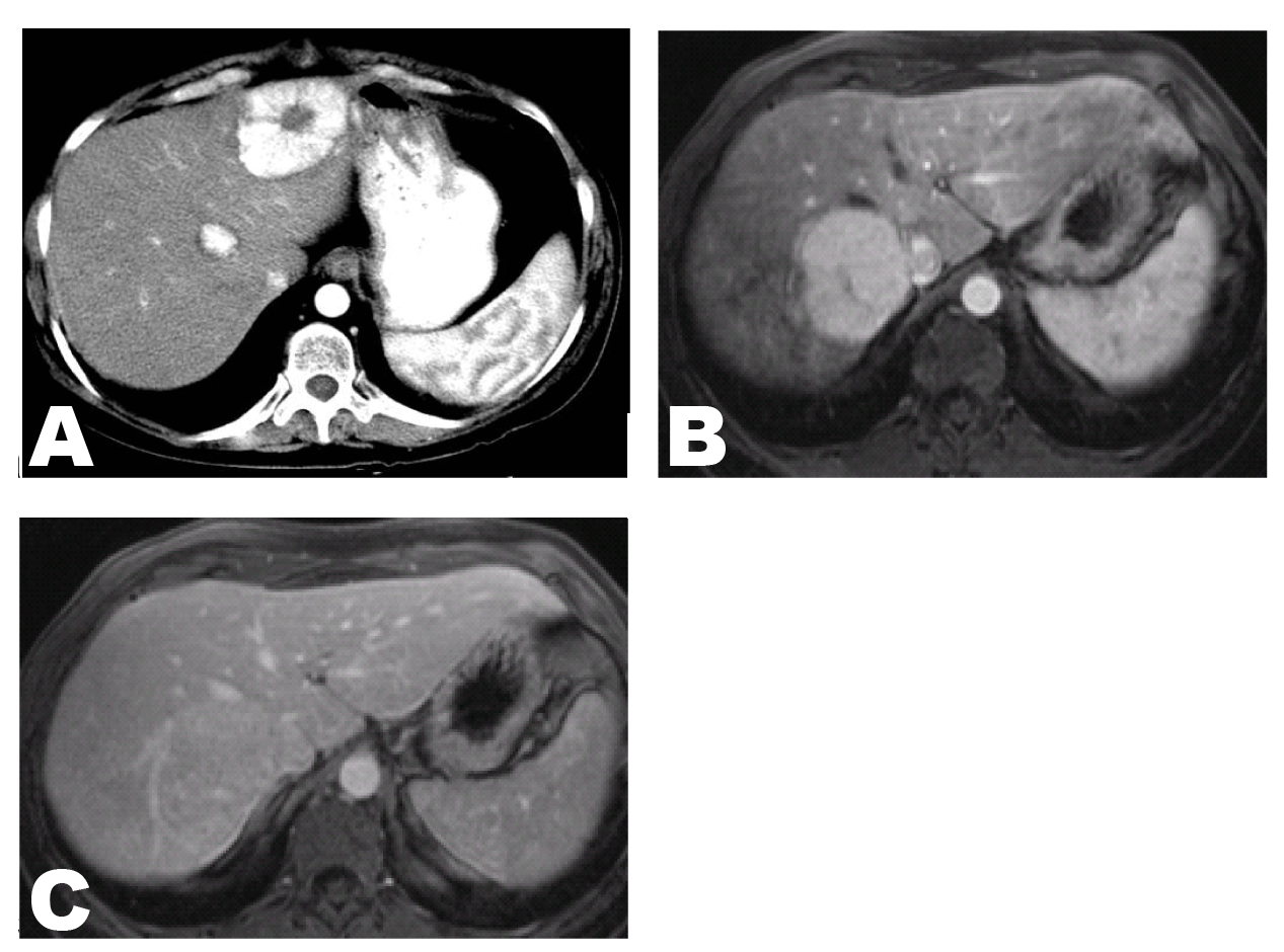
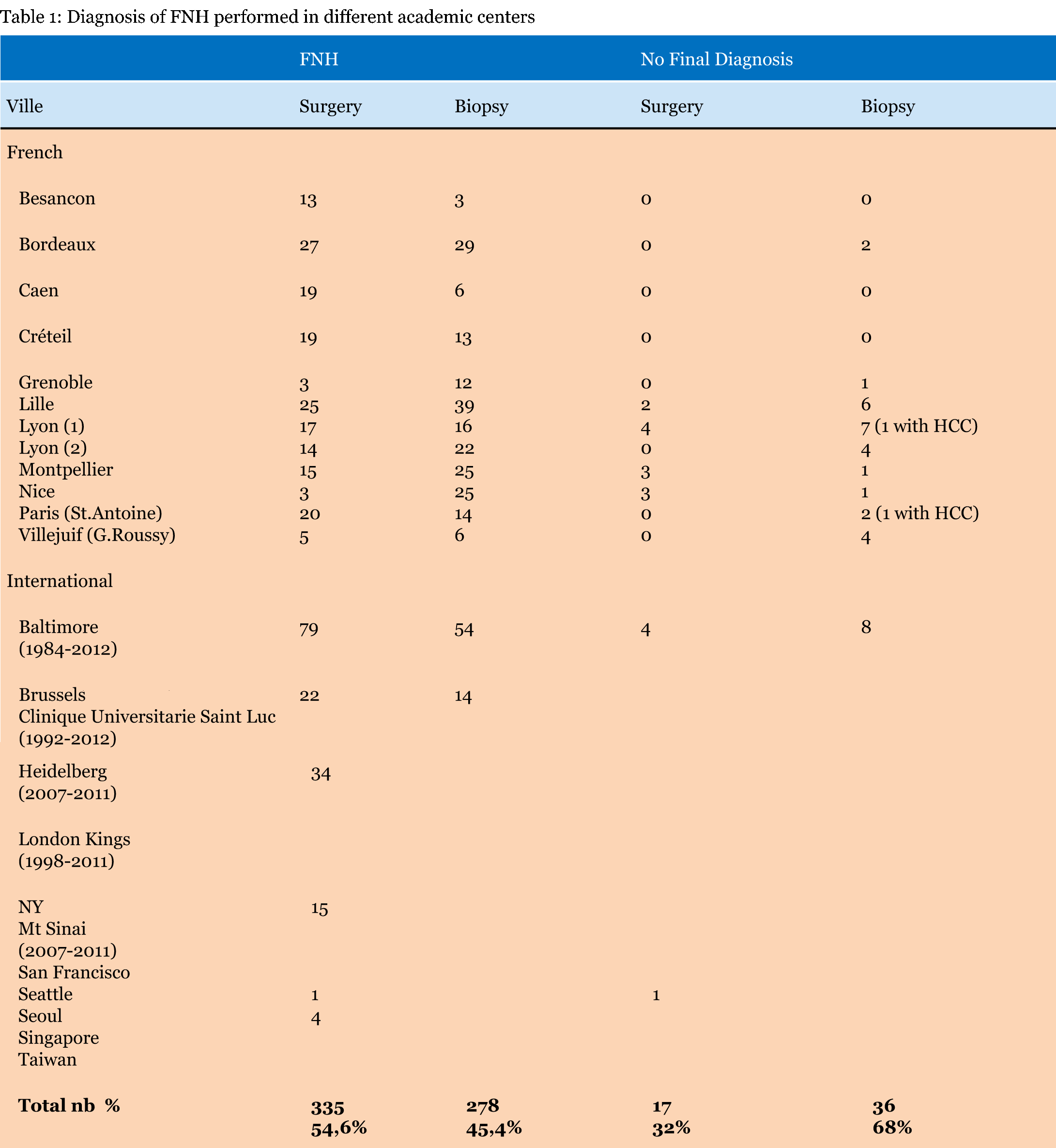
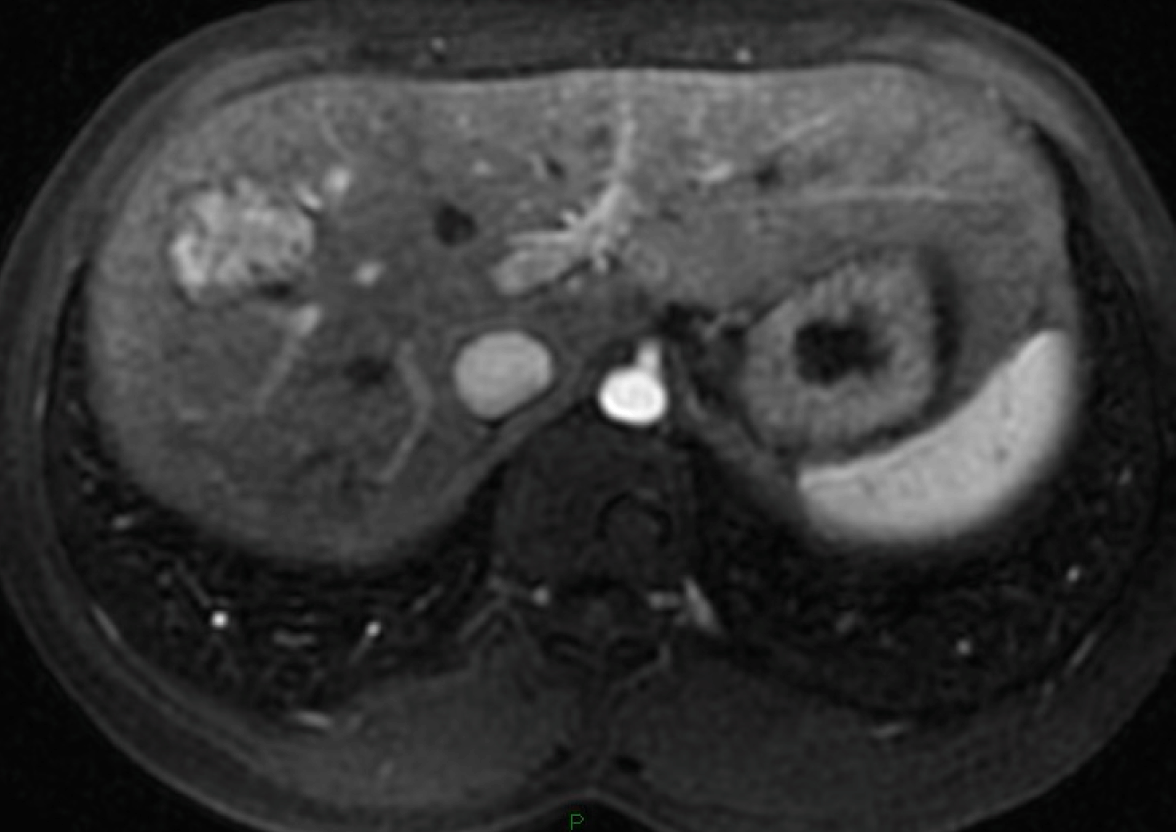
This is the second most prevalent benign liver lesion (after hemangioma), with a woman preponderance, in 80–90% of the cases, in third or fourth decade and a global incidence of about 0.6–3% of the general population. It is no identifiable etiologic factor. It is, however, associated with a conditions having local or systemic vascular anomalies. Focal nodular hyperplasia (FNH) has a demographic variation with a male and children of either gender preponderance in countries where oral contraceptives use has been less prevalent (i.e., China). There is not the impact on size variation from oral contraceptives use and from pregnancy and is not indispensable, though recommended, to stop oral contraceptive use. [3] Most of the patients are not symptomatic and the diagnosis is made incidentally during surgery, autopsy or imaging procedures for others symptoms. The liver function tests is normal complications, as the rupture, bleeding or malignant transformation, although rare, are described in literature. [4] The diagnosis of FNH can be made using imaging techniques in 90% of cases in experienced centers. [5] [6] Today, surgery and biopsy are performed in some academic centers in Europe and United States for diagnosis of FNH. Contrast-enhanced ultrasonography (CEUS) is the first modality of choice for FNH:FNH typically shows arterial increased enhancement, very marked in the first few seconds. Centrifugal (70%) or eccentric (30%) enhancement through one afferent correspondingly situated arteries is a diagnostic pointer. In the portal venous phase FNH shows at least low-grade increased enhancement in about 95% of cases. The centrifugal filling sign is very useful for diagnosis of FNH. [7] [8] Recently in 2013, Wang et al. from the University of Guangzhou (Republic of China) published their experience with CEUS in 85 patients with 85 histological proven FNH. Enhancement, centrifugal filling, spoke-wheel arteries were reviewed and correlated with the size. Forty-seven other focal liver lesions with contrast-enhanced computed tomography (CECT) were randomly selected for comparison of diagnostic with CEUS. The results confirm that the CECT have similar diagnostic performance for FNH (Figure 1A) and CEUS should be the first imaging technique for the diagnosis of FNH. [9] The magnetic resonance imaging (MRI) scan has the advantage of being a non-radiating technique and has the excellent contrast resolution. In MRI, a typical FNH is hyperintense in the arterial phase and isointense before contrast and in the portal venous phase (Figure 1B-C). In 89% cases, before contrast FNH is hyperintense to isointense on T2 and isointense to hypointense on T1. [10] [11] [12] Half of FNH has a central scar, slightly hyperintense on T2 and with late gadolinium-contrast enhancement. However, the choice between CEUS and MRI depends upon the experience and local availability. In less than 10% of cases, the differential diagnosis of FNH, HCA and HCC cannot be solved by imaging alone. A biopsy can resolve most of these problem cases, with standard and/or immunohistochemical staining. [13] [14] The histological diagnosis of FNH requires two main criteria: the lesion must be composed of benign-appearing hepatocytes and must be supplied by altered portal tracts. The risk of complications as hemorrhage and malignant transformation is absent and surgery is not indicated. However, it is still performed for organ compression, compression of liver vessels and biliary tract, pain and especially if doubt about the nature of the tumor in the absence of histological diagnosis. To date, the unavailability of guidelines hampers the follow-up of these lesions. Regarding the management and treatment of FNH, we can summarized as follow: If the patient is female with typical FNH on imaging, normal liver tests and no medical history of cancer, the diagnosis of FNH is considered and the biopsy is not necessary. If the patient is a male, biopsy is proposed. In the case of atypical FNH on imaging biopsy is necessary. [15] FNH is often asymptomatic and the surgery is not indicated even if the large lesion. However, the patients with a large lesions can to develop abdominal pain or compression of adjacent structures and liver resection may be indicated. Laparoscopic enucleation without coverage of the resection margins is a contemporary approach. Relating to surveillance Cherqui D [16] published a short report on clinical management of benign liver cell tumors and it is proposed for FNH monitoring by a MRI to six and twelve months,then MRI or CEUS each year for three years; after to stop monitoring. [16] | ||||||
|
| ||||||
|
Hepatocellular Adenoma
| ||||||
|
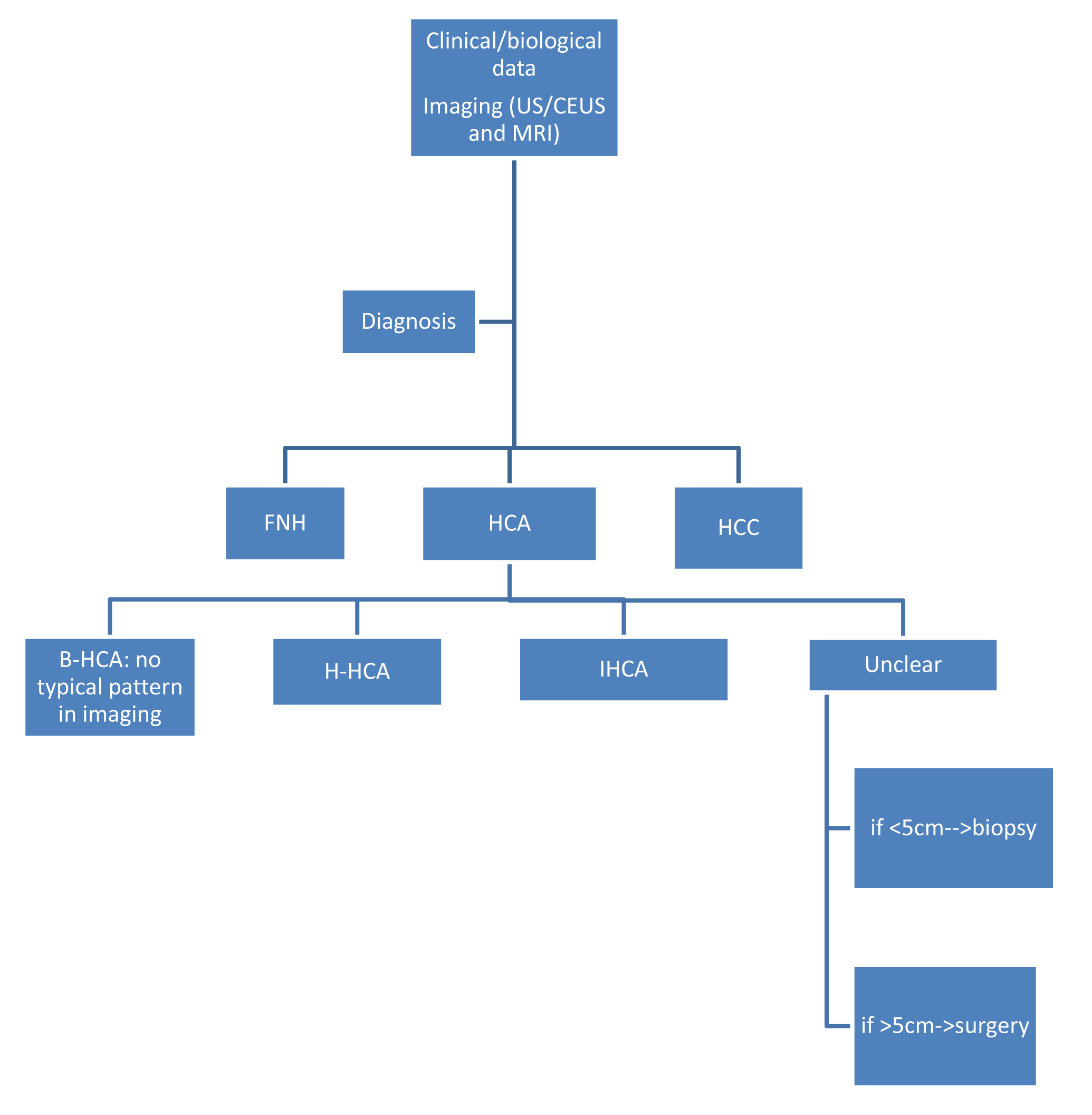
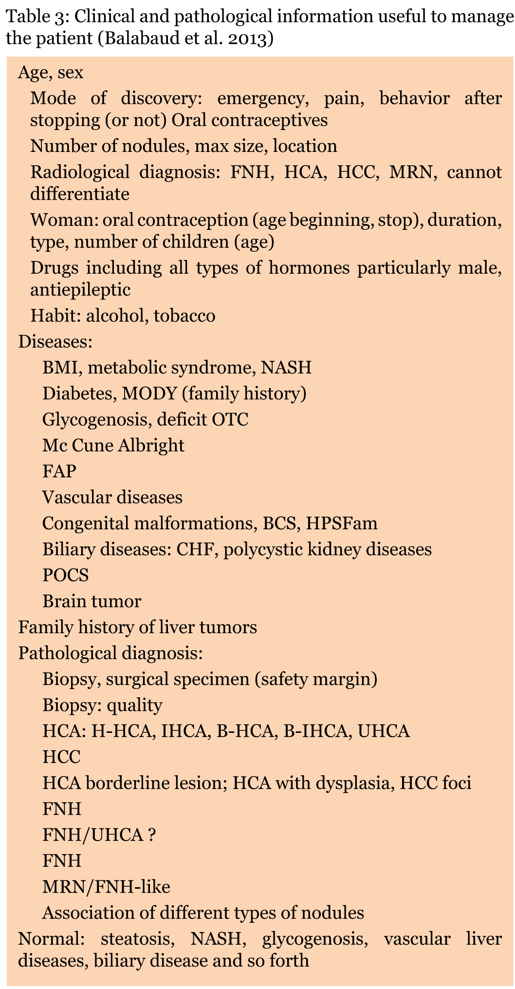
This is an uncommon benign liver lesion arising from monoclonal proliferation of hepatocytes with a female preponderance before menopause and after a long- term use of oral contraception. It is associated with the duration and type of oral contraceptives usage. Other risk factors include: glycogen storage diseases and androgen intake. Complications as hemorrhage and more rarely malignant transformation is reported. [17] [18] [19] [20] The major classification [21] of HCA according to molecular site [21] [22] [23] [24] [25] [26] divide patients with hepatocellular adenoma (HCA) in four subgroups according to their genotype/phenotype. (Table 2) The first subgroup: hepatocyte nuclear factor 1 a (HNF1 a),as the first driver gene inactived by mutation in hepatocellular adenomas and it occurs in almost exclusively all cases in females. [27] In HCA tumor cells with mutations of HNF1 a, we described complete HNF1 a inactivation by mutation of both alleles in 35–45% of the cases and by immunohistochemitry there is a complete absence of liver fatty acid binding protein (L-FABP), specific of this subtype. (Table 2)[28] The second subgroup:mutations activating β-catenin are described in 10–15% of HCA. (Table 2) CTNNB1, the gene coding for β-catenin, is the most mutated oncogene in HCC (from 20–40% of the cases). The β-catenin subtype is often associated with conditions such as glycogenosis and male hormone administration and it occurs in almost exclusively in man. It is very important that HCA with activating mutation of β-catenin have a high risk of malignant transformation in HCC compared to other subtypes. [28] It should know that distinguishing HCA from well-differentiated HCC developed on normal liver could be challenging to diagnosis. Consequently, all patients identified with a mutation of β-catenin should be considered for liver resection to avoid the risk of malignant transformation. The third subgroup: Hepatocellular adenomas with inflammatory features (IHCA). Inflammatory HCA presented a cytoplasmic over-expression of SAA and CRP, two proteins of the acute phase of inflammation, in the tumor hepatocytes. (Table 1)[29] IHCA is associated frequently with high alcohol consumption and obesity, two conditions associated with chronic cytokine production and it occurs often in woman. [30] The fourth subgroup: Ten percent of HCA no identifiable etiologic factor. (Table 2) Pratical guidelines for the diagnosis of HCA subtypes are summarized in Table 3. Once the diagnosis of liver tumor is made, the patient is often referred to a surgeon. The identification of subtypes is one of the key factors among others that need to be collected. (Figure 2) (Figure 3) There are neither specific guidelines nor a standardized therapeutic approach available to date to its management. [31] [32] [33] However, there is a consensus according to the HCA >5 cm should be resected if they have not regressed after stopping oral contraceptives, particularly those at a high risk of malignant transformation (β-HCA and β-IHCA). (Figure 4) Some surgeons prefer that all HCA should be removed particularly, if they are easily accessible laparoscopically. [34] [35] [36] Balabaud C et al. from the University of Bordeaux, reviewed their experience and suggested the management of HCA subtypes <5 cm as it is summarized in (Table 3) (Figure 5)[31] They proposed in female patients, if size increases after follow-up and to stop oral contraceptive, resection if the size of lesion is >5 cm, though the resection should be considered for nodules <5 cm (in the 3–4 cm range) and resection independently of the size in the male. A biopsy is very performed in patients at risk of malignant transformation such as young woman/metabolic or vascular disorders. In presence of patients with H-HCA associated to abnormal genetic counseling resection should be considered independently of the size. Future development will be based on imaging techniques, molecular data including chromosomal abnormalities, in the hope of being able to combine molecular, radiological and clinical data. | ||||||
| ||||||
| ||||||
| ||||||
| ||||||
| ||||||
| ||||||
|
| ||||||
| ||||||
|
Conclusion
| ||||||
|
In future, we will be able to propose a specific guideline which can be a multidisciplinary approach (hepatologist, surgeon, radiologist and pathologist) towards the overall management of these diseases and is key factor for a good outcome. | ||||||
|
Abbreviations
| ||||||
|
US: Ultrasound | ||||||
|
References
| ||||||
| ||||||


|
[HTML Abstract]
[PDF Full Text]
|
|
Author Contributions
Elisa Palladino – Substantial contriutions to conception and designs, Acquisition of data, Drafting the article, Revising it critically for important intellectual content, Final approval of the version to be published Daniele Sommacale – Substantial contriutions to conception and designs, Acquisition of data, Drafting the article, revising it critically for important intellectual content, Final approval of the version to be published Renaud Siboni – Substantial contriutions to conception and designs, Acquisition of data, Drafting the article, Revising it critically for important intellectual content, Final approval of the version to be published Christine Hoeffel – Substantial contriutions to conception and designs, Acquisition of data, Drafting the article, Revising it critically for important intellectual content, Final approval of the version to be published Christian Lechner – Substantial contriutions to conception and designs, Acquisition of data, Drafting the article, Revising it critically for important intellectual content, Final approval of the version to be published Tullio Piardi – Substantial contriutions to conception and designs, Acquisition of data, Drafting the article, Revising it critically for important intellectual content, Final approval of the version to be published Reza Kianmanesh – Substantial contriutions to conception and designs, Acquisition of data, Drafting the article, Revising it critically for important intellectual content, Final approval of the version to be published |
|
Guarantor of submission
The corresponding author is the guarantor of submission. |
|
Source of support
None |
|
Conflict of interest
Authors declare no conflict of interest. |
|
Copyright
© 2014 Elisa Palladino et al. This article is distributed under the terms of Creative Commons Attribution License which permits unrestricted use, distribution and reproduction in any medium provided the original author(s) and original publisher are properly credited. Please see the copyright policy on the journal website for more information. |
|
|
|
About The Authors
| |||
| |||
| |||
| |||
| |||
| |||
| |||
| |||
