 |
|
Case Report
|
| Avulsion of the common bile duct and ampulla of Vater after blunt abdominal trauma: Case report and review of the literature |
| Ayman Zaki Azzam1 |
|
1Lecturer of General Surgery, Department of General Surgery, Alexandria University, Alexandria, Egypt.
|
|
Article ID: 100001IJHPDAZA2011 doi:10.5348/ijhpd-2011-1-CR-1 |
|
Address correspondence to: Ayman Zaki Azzam Lecturer of General Surgery, Faculty of Medicine, Alexandria University, Alexandria Egypt Liver Transplant Surgeon, King Faisal Specialist Hospital and Research Center, MBC: 72, P.O.Box 3354 Riyaddh 11211 South Arbia Phone: 966-1-464-7272, ext 39474 Fax: 966-1-442-4817 |
|
[HTML Abstract]
[PDF Full Text]
|
| How to cite this article: |
| Azzam AZ. Avulsion of the common bile duct and ampulla of Vater after blunt abdominal trauma: Case report and review of the literature. International Journal of Hepatobiliary and Pancreatic Diseases 2011;1:1-5. |
|
Abstract
|
|
Introduction:
Extrahepatic bile ducts injuries from blunt abdominal trauma are very rare. The criticality and the difficulty of these injuries are increased by the duodenal wall and the main pancreatic duct injuries. These injuries therefore require special consideration. Diagnosis is always difficult and late and depends on suspicion. Proper management depends on the efficiency of the center, the surgeon and the severity of the injury.
Case Report: A 28-years-old male presented to the emergency unit complaining of falling from motor cycle. The patient was stable. Urgent ultrasound revealed mild intraperitoneal collection. Computerized tomography with contrast showed the same finding with left sided pleural effusion and fracture fifth rib. The patient's condition began to deteriorate with time. Immediate exploration revealed complete avulsion of the CBD from the duodenum. Exteriorization of the CBD to outside the abdomen through the skin was done. The diagnosis of avulsion of the CBD from the sphincter of Oddi was a retrospective diagnosis. Conclusion: Simple maneuvers can be performed that will not harm the patient and may be of great benefit. In the present case, exteriorization of the CBD to outside by a simple urinary catheter gave us the chance to have an accurate postoperative diagnosis which was not available at the time of the operation. Drainage of the pancreatic and biliary secretions outside the abdomen with no added morbidities also gave us the chance and the time needed to treat other associated complications like respiratory problems. | |
|
Keywords
Common bile duct, Ampulla of Vater, Blunt abdominal trauma
| |
|
Introduction
| ||||||
|
Avulsion of the extrahepatic bile ducts from blunt abdominal trauma is an infrequently encountered condition [1]. The complexity of these injuries is increased by the degree of involvement of the duodenal wall and the main pancreatic duct. The injuries that include the ampullary area are rare, and therefore require special consideration. Only few reports of these lesions have appeared in English literature. Failure to recognize the clinical manifestations of these uncommon injuries results in delayed diagnosis. [2] Very often the lesion is not identifiable until the signs of jaundice and biliary ascites occur. When the diagnosis is late the corner stone of treatment is biliary diversion and definitive repair after complete resolution of sepsis with a choledochojejunostomy [3]. | ||||||
|
Case Report
| ||||||
|
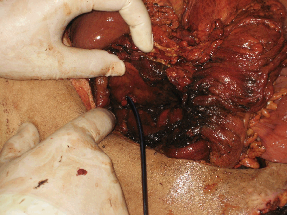
A 28-year-old male patient, opiates addict, presented to the emergency unit, General Surgery Department, Faculty of Medicine, Alexandria University, Egypt, complaining of fall from motor cycle. A transverse contused wound in the abdomen about 30 cm below the umbilicus was present. The patient was hemodynamically stable, the pulse was 78 beats/min, blood pressure was 115/70 mmHg and the respiratory rate was 22/min. The abdomen was lax and intestinal peristalsis was heard. Urgent ultrasonogram (USG) was done and revealed mild free intraperitoneal collection. Computerized tomography (CT) scan with contrast showed the same finding with left sided pleural effusion and fracture of fifth rib. Intravenous fluids were started. The anterior abdominal wall wound was closed by direct sutures after thorough cleaning. The vital signs were monitored regularly. The patient was planned to be put on conservative management and was transferred to the ward. Repeated USG of the abdomen was done daily for the next five days without any interval changes. The patient was vitally stable during that period. Follow up CT scan showed free intraperitoneal collection in the Morrison's pouch, perisplenic, right and left paracolic and pelvic regions. The CT through the lower chest revealed bilateral, moderate pleural effusion, more on the right side with consolidating collapse of the posterior segments of both lower lobes. Cardiothoracic surgeon was consulted, who decided to put a right intercostal tube. The patient's condition began to deteriorate slowly with time. After seven days follow-up in the ward, the patient developed mild fever. Abdomen was slightly distended, became tender with rebound tenderness and silent. A decision was taken to do immediate exploratory laparotomy. A midline incision was made. The abdomen was full of bile, which was staining the small and large bowels. After evacuation of about three liters of bile from the abdomen, meticulous exploration of the abdomen revealed complete avulsion of the CBD from the duodenum. The duodenum was also avulsed from the head of pancreas, and both were avulsed from the retroperitoneal portion. The duct was cannulated with an 8 Fr urinary catheter to detect its pathway (Figure 1). The distal opening in the duodenum was searched for but unfortunately it was not found. Introduction of the nasogastric tube to the end of the second portion of the duodenum and closure of the duodenum by non-crushing clamp followed by
Figure1: Intraoperative view showing avulsed duodenum from the head of pancreas and the urinary catheter passing through the proximal avulsed end of CBD.
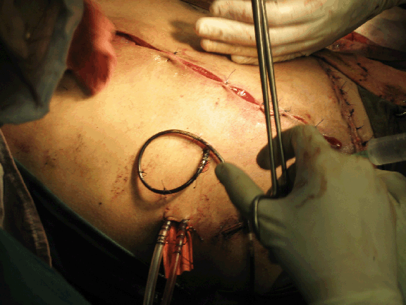
injection of normal saline through the nasogastric tube to the duodenum did not reveal any leak. The site of the opening of the sphincter of Oddi was not found. Complete exploration of the abdomen did not reveal any other lesion. The liver was also intact. Due to the presence of unhealthy tissues and the presence of abdominal infection, the plan was exteriorization of the CBD to outside the abdomen through the skin. This was performed by inserting 8 Fr catheter through the skin, then behind the duodenum, then through the CBD and was transfixed to the CBD by vicryl suture 4/0 and then fixed to the skin by silk sutures 2/0 in multiple sites to prevent fall of the catheter. Peritoneal lavage and insertion of a rubber drain in the Morrison's pouch and multiple drainage tubes in the pelvis and subdiaphragmatic space was done (Figure 2).
| ||||||
|
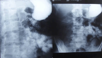
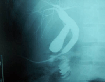
The patient was transferred to the ICU. He was put on ventilator. He was suffering from withdrawal symptoms of addiction in the form of agitation, anxiety, depression, sweating, increased salivation and aggressive behavior. The course of the patient in the ICU was bad in the start because of the withdrawal symptoms of the addiction. It was managed by the ICU physician by supportive care and opiates substitutes. After extubation, the patient developed respiratory problems in the form of tachypnea, dyspnea and difficulty in breathing (respiratory rate > 50/min). Intercostal tube was inserted in the right side on the 2nd day and another one was inserted in the left side on the 5th day due to accumulating pneumothorax. The patient was intubated again and put on ventilator for two days, then extubated and put on face mask. The daily amount of the draining fluid through the percautaneous catheter was about 1000-1500cc and from the rubber drain about 500 cc. Follow up USG Figure 4: Gastrographin follow-through showing free flow of the dye with non visualization of the duodenum without definite evidence of contrast leak. The surgical rubber drainage and catheter are seen at the right hypochondrial region Figure 5: Follow-up Percautaneous cholangiogram through the external catheter after 5 months, showing dilatation of the previously avulsed CBD. The bowel loops are opacified with the contrast?? abdomen revealed minimal free intraperitoneal fluid collection in the subhepatic, perisplenic and pelvic regions. Gastrografin follow through was done and revealed free flow of the dye in the intestine, with non visualization of the duodenum without definite evidence of contrast leak (Figure 4). During the ICU admission, the peristalsis regained and the bowel motion was restored. The drains were removed and the patient started oral feeding. Follow-up CT of the chest revealed minimal pneumothorax on the right side, and improvement in the basal lung consolidation on the left side. Both intercostal tubes were removed. The patient spent about 15 days in the ICU during which his general condition and laboratory work began to improve progressively. Serum amylase level was high after ICU admission and began to decrease to around normal figure by the end of ICU stay, but amylase levels from the drain remains in the range of 950,000-1200,000 U/ml. The patient was transferred to the ward and stayed for about one week, during which his condition improved much. He was then discharged with the percautaneous catheter tube in the CBD with about 1500 cc/day. The plan was for another operation after improvement of the general condition and after doing the proper evaluation of the patient's condition. After five months of follow up, the general condition of the patient was good. He regained his normal body weight and restored his renal and liver functions. He was admitted for second operation to repair the CBD and resume the biliary and pancreatic drainage. Percautaneous cholangiography through the external catheter on the skin was done and revealed dilatation of the previously avulsed CBD and the catheter appearing coming out from it. The bowel loops are opacified with the contrast (Figure 5). | ||||||
| ||||||
|
| ||||||
|
| ||||||
|
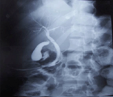
The diagnosis of avulsion of the CBD from the sphincter of Oddi was a retrospective diagnosis after performing cholangiography through the external catheter on the skin which revealed the presence of both the bile and pancreatic ducts in the proximal opening of the avulsed end of the common bile duct (Figure 3). Also, the amylase content of the secretion coming from the percautaneous catheter put in the CBD contains more than 1,200000 U/ml. | ||||||
|
| ||||||
|
| ||||||
|
| ||||||
|
| ||||||
|
There was a debate on how to manage the biliary drainage for that patient and whether he will need Whipple's procedure or not. After opening the abdomen, the adhesions were extensive. Adhesiolysis was done and the CBD was found, guided by the external catheter which was previously put. The external catheter had already paved a track from the CBD to the skin, behind the duodenum. The CBD was found to be dilated more and acquired thick wall which make it easier to be anastomosed with the jejunum. After dissection, the fistulous track behind the duodenum was separated and the CBD was anastomosed to the jejunum end to side over a stent and drains were inserted. Cholecystectomy was done. The patient spend about one week in the hospital and then discharged in a good condition after eating and drinking and after performing the laboratory investigations. After one month he came for follow up. The stent was removed. His general condition was very good, and was advised for continuous follow up. | ||||||
|
Discussion
| ||||||
|
The exact nature of the force necessary to cause injury to the extrahepatic bile ducts (EBD) is unknown although impingement and compression of the ductal system on the vertebral column, [4] external compression of the gall bladder with transmitted rise in intraductal pressures resulting in "blowout of the duct", [5] and laceration of the ductal system at the junction of its fixed and mobile portions, [6] [7] may explain EBD injuries. The exact pathophysiology still remains unclear, but a combination of the above theories may be responsible for EBD injuries. Complete and partial lacerations occurring in all portions of EBD have been previously described. Review of literature revealed the rarity of EBD lesions. Rydell reported transected common bile duct in 25 cases and lacerated common bile duct in 31 cases. [8] Since that time, avulsion of the Ampulla of Vater [9][10][11] as well as several other reports on EBD injuries have been added. [12] [13] [14] [15] Total transection of the duct can be repaired by primary end-to-end anastomosis if the case is freshly diagnosed with no infection and the distal end of the duct is identified and healthy. Primary choledochoduodenostomy or Roux-en-Y choledochojejunostomy are other options if the distal end is not identified or there is fear of postoperative stricture. Injuries to the common bile duct together with or in the vicinity of the main pancreatic duct are the most severe and present the most challenging clinical problem. In the present case there was laceration and avulsion of the head of the pancreas from the C shape of the duodenum and also the duct was avulsed completely from the duodenum with failure to detect the site of sphincter of Oddi in the duodenum. The avulsion of the pancreatic duct with the bile duct was a possibility with the absence of operative cholangiogram facility in the emergency OR room in our University. Also, the presence of infection made the primary repair impossible. The decision to exteriorize the CBD and close the abdomen for 2nd look after 24-48 hours was to assess the viability of the pancreas and the duodenum and to do percautaneous transcatheter cholangiogram to make an exact diagnosis and to decide the most suitable plan for management. Fortunately, the patient improved as regard the abdominal symptoms but his general condition deteriorated due to the chest condition. So the exteriorization of the CBD that was done took less time and gave us the chance to assess the magnitude of the damage and also to give us a chance to see whether the pancreas and duodenum will resolve or not. In the present case, although there was a complete avulsion of the pancreas from the duodenum, they both resolved completely without any need for further intervention which gave us the chance to manage other morbidities of the patient and postpone the problem of CBD for future intervention. In summary, reports about CBD injuries are rare, and the reports of avulsion of the Ampulla of Vater are extremely rare. Moreover, in difficult circumstances, such as hospitals in undeveloped countries without all the facililities and the equipments to diagnose and manage the problem, or, if the diagnosis is made late and after development of abdominal infection (biliary peritonitis), we have to do our best to save the patient's life by simple maneuvers that will not harm the patient and may be of great benefit. In the present case, exteriorization of the CBD to outside by a simple urinary catheter gave us the chance to have an accurate postoperative diagnosis which was not available at the time of the operation. Also, drainage of the pancreatic and biliary secretions outside the abdomen with no added morbidities, also, this gave us the chance and the time needed to treat other complications associated like respiratory problems as in our case. Postoperative follow up cholangiography was also very easy to follow the patients condition and to decide the future plans for biliary reconstruction. | ||||||
|
Conclusion
| ||||||
|
Simple maneuvers can be performed that will not harm the patient and may be of great benefit. In cases of CBD injuries, exteriorization of the CBD to outside by a urinary catheter is simple, easy to perform procedure and can give the chance to make an accurate postoperative diagnosis which may be not available at the time of the operation. Drainage of pancreatic and biliary secretions outside the abdomen, also, can give the chance and the time needed to treat other associated complications. | ||||||
|
References
| ||||||
| ||||||

| [HTML Abstract] [PDF Full Text] |
|
Author Contributions:
Ayman Zaki Azzam - Substantial contributions to conception and design, acquisition, analysis and interpretation of data, Drafting the article, revising it critically for important intellectual content, Final approval of the version to be published |
|
Guarantor of submission:
The corresponding author is the guarantor of submission. |
|
Source of support:
None |
|
Conflict of interest:
Author declares no conflict of interest. |
|
Copyright:
© Ayman Zaki Azzam 2011; This article is distributed the terms of Creative Commons attribution 3.0 License which permits unrestricted use, distribution and reproduction in any means provided the original authors and original publisher are properly credited. (Please see Copyright Policy for more information.) |
|
|