|
Case Report
Pancreaticopleural fistula secondary to chronic calcific pancreatitis presenting with massive left-sided pleural effusion: A case report
1 MD, Radiology Specialist, Radiology Department, Sohar Hospital, Sohar, North Al Batinah, Sultanate of Oman
2 MD, Consultant Radiologist, Radiology Department, Sohar Hospital, Sohar, Sultanate of Oman
3 Specialist Radiology, Radiology Department, Sohar Hospital, Sohar, North Al Batinah, Sultanate of Oman
Address correspondence to:
Jamila Omar Abdalla
MD, Specialist Radiology, Radiology Department, Sohar Hospital, Sohar, North Al Batinah,
Sultanate of Oman
Message to Corresponding Author
Article ID: 100113Z04JA2026
Access full text article on other devices

Access PDF of article on other devices


How to cite this article
Abdalla JO, Mohamed RMI, Selim YAR, Awadelkreem AO, Al-Jabri FSA. Pancreaticopleural fistula secondary to chronic calcific pancreatitis presenting with massive left-sided pleural effusion: A case report. Int J Hepatobiliary Pancreat Dis 2026;16(2):22–27.ABSTRACT
Introduction: Pancreaticopleural fistula (PPF) is an uncommon complication of pancreatitis that often presents with respiratory symptoms, making its pancreatic origin difficult to recognize initially.
Case Report: A 41-year-old man presented with shortness of breath and severe left-sided chest pain. He reported no abdominal symptoms, and abdominal examination revealed a soft abdomen. Chest radiography demonstrated a left-sided pleural effusion, which markedly increased over the following three days, prompting further evaluation with contrast-enhanced computed tomography (CT). Contrast-enhanced CT demonstrated chronic calcific pancreatitis with pancreatic atrophy and ductal dilatation, together with a long peripherally enhancing fistulous tract extending from the pancreatic duct to the left pleural cavity. Pleural fluid analysis showed markedly elevated amylase levels, consistent with PPF. Subsequent review of the patient’s medical records revealed a history of hemorrhagic pancreatitis. The patient later underwent surgical treatment, including distal pancreatectomy, excision of the PPF, splenectomy, and cholecystectomy. His postoperative recovery was uneventful.
Conclusion: Pancreaticopleural fistula should be suspected in patients presenting with massive unilateral pleural effusion, especially when respiratory symptoms are prominent and abdominal symptoms are mild or absent. Contrast-enhanced CT can demonstrate the fistulous tract, helping to confirm the diagnosis and plan appropriate management.
Keywords: Chronic calcific pancreatitis, Computed tomography, Pancreaticopleural fistula, Pleural effusion
Introduction
Pancreaticopleural fistula (PPF) is a rare complication of pancreatic disease, most commonly associated with pancreatitis and pancreatic duct disruption. Pancreatic secretions can extend into the thoracic cavity, resulting in a large or recurrent pleural effusion [1],[2].
Diagnosis can be challenging because respiratory symptoms often predominate, while abdominal symptoms may be mild or absent. This may delay recognition of the pancreatic origin of the pleural effusion [1],[3].
Computed tomography (CT) and magnetic resonance cholangiopancreatography (MRCP) are useful for identifying the underlying pancreatic abnormality and demonstrating the fistulous tract, while markedly elevated pleural fluid amylase provides strong biochemical support for the diagnosis [1],[2].
We report a case of pancreaticopleural fistula secondary to chronic calcific pancreatitis presenting with left-sided pleural effusion, in which the fistulous tract was clearly demonstrated on contrast-enhanced CT.
Case Report
A 41-year-old man with a history of smoking, but no known history of diabetes mellitus or hypertension, presented with a 3-month history of mild productive cough, followed by four days of intermittent low-grade fever, shivering, shortness of breath, and severe left-sided chest pain. He denied sweating or hemoptysis. He reported no associated abdominal symptoms.
Further history revealed that the patient had smoked 6–7 cigarettes daily for 20 years (approximately 6–7 pack-years) and denied alcohol consumption or other known chronic medical conditions. Subsequent review of his previous medical records revealed a history of hemorrhagic pancreatitis in 2017, complicated by a retrogastric hematoma (8 × 4 × 5 cm) and intra-abdominal fluid collections, requiring laparoscopic drainage of a lesser sac collection. His serum calcium level at that time was 8.2 mg/dL. Triglyceride levels and investigations for other etiologies were unavailable, and the underlying cause of chronic pancreatitis remained uncertain. On admission, he was hemodynamically stable, with a blood pressure of 126/70 mmHg and oxygen saturation of 97% on room air. Abdominal examination revealed a soft, non-distended abdomen.
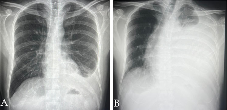
Laboratory tests showed an erythrocyte sedimentation rate (ESR) of 40 mm/h and a C-reactive protein (CRP) level of 19.82 mg/L (reference range: 0–5 mg/L). Initial chest radiography demonstrated a left-sided pleural effusion (Figure 1A). Tuberculosis and malignancy were considered in the differential diagnosis. A follow-up chest radiograph obtained three days after admission demonstrated a marked increase in the left pleural effusion (Figure 1B). This finding prompted further assessment with contrast-enhanced chest CT.
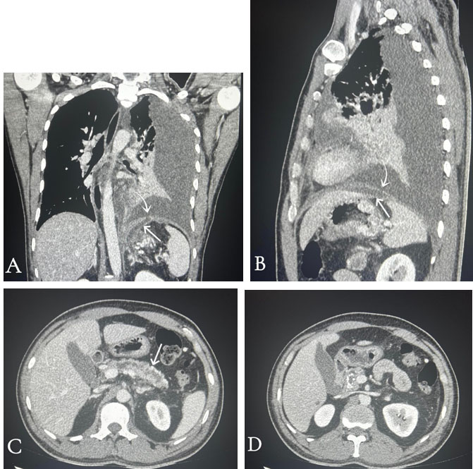
Contrast-enhanced CT of the chest showed a massive left pleural effusion. The included upper abdominal images revealed pancreatic atrophy, dilatation of the main pancreatic duct, and multiple calcifications, in keeping with chronic calcific pancreatitis. A long peripherally enhancing fistulous tract was identified arising from the pancreatic duct at the body–tail junction and extending into the left pleural cavity through a direct diaphragmatic defect, rather than through the aortic or esophageal hiatus, consistent with pancreaticopleural fistula (Figure 2). A small pancreatic pseudocyst was also noted. A left-sided intercostal chest drain was inserted, initially draining approximately 1500 mL of pleural fluid. Subsequent recorded outputs were 260 mL and 20 mL over 24 hours. Pleural fluid sampling was performed after CT imaging, and subsequent biochemical analysis revealed markedly elevated amylase levels.
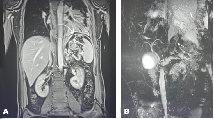
Pleural fluid analysis showed a markedly elevated amylase level of 4407.60 U/L, whereas the serum amylase level was 107.30 U/L (reference range: 22–80 U/L), supporting the diagnosis of pancreaticopleural fistula. Abdominal magnetic resonance imaging (MRI) with MRCP was then performed, demonstrating the pancreatic duct abnormality and fistulous communication, in agreement with the CT findings (Figure 3).
Endoscopic retrograde cholangiopancreatography (ERCP) was planned for further assessment and potential treatment; however, the patient declined the procedure, and ERCP was not performed. His clinical condition improved during hospitalization. He later underwent surgical management without prior endoscopic intervention, including distal pancreatectomy, excision of the pancreaticopleural fistula, splenectomy, and cholecystectomy.
The patient remained hospitalized for two weeks following surgery and had an uneventful postoperative recovery. Postoperative chest radiography reportedly demonstrated resolution of the pleural effusion; however, the images were not available for review. At the one-month follow-up, the patient was clinically well. He has since returned to work and remains in good general health.
Histopathological examination of the resected distal pancreas revealed hemorrhage and fibrosis with benign pancreatic cells. The spleen showed chronic passive congestion, while the gallbladder showed chronic congestion. No evidence of malignancy was identified in the examined tissues.
Discussion
Pancreaticopleural fistula is an uncommon complication of pancreatitis, usually caused by disruption of the pancreatic duct or leakage from a pancreatic fluid collection. Pancreatic secretions can pass through the retroperitoneum and reach the thoracic cavity via diaphragmatic openings or direct extension across the diaphragm. Chronic pancreatitis is the most frequent underlying cause, but PPF can also develop following acute pancreatitis or pancreatic trauma [1],[2],[4].
Respiratory symptoms are often more prominent than abdominal symptoms, which may be minimal or absent. Common presentations include dyspnea, cough, chest discomfort, and recurrent pleural effusion, often raising suspicion of a primary thoracic condition [1],[3],[5]. In our patient, fever, cough, shortness of breath, and massive left-sided pleural effusion initially raised concern for tuberculosis or malignancy. Contrast-enhanced CT subsequently revealed findings suggesting a pancreatic origin.
Diagnosis of pancreaticopleural fistula is based on both imaging and biochemical findings. Markedly elevated pleural fluid amylase is an important clue and should raise suspicion of a pancreatic source in patients with massive or recurrent pleural effusion [1],[2]. Imaging is used to assess the pancreas, pancreatic duct, and the fistulous tract when visible [2].
Magnetic resonance cholangiopancreatography allows non-invasive evaluation of the pancreatic duct and can help identify the fistulous communication. It is generally more sensitive than CT for demonstrating pancreatic duct abnormalities and fistulous tracts. Computed tomography is also valuable, especially in cases where a pancreatic origin is not initially suspected [2].
In our patient, contrast-enhanced CT clearly showed chronic calcific pancreatitis, dilatation of the main pancreatic duct, and a long fistulous tract extending to the left pleural cavity. The tract was well visualized on CT before biochemical confirmation. Magnetic resonance cholangiopancreatography provided findings similar to those seen on CT and supported the diagnosis.
Management of pancreaticopleural fistula depends on the pancreatic duct abnormality, fistula anatomy, and the patient’s clinical condition. Conservative treatment, including pleural drainage and measures to reduce pancreatic secretion, may be used in selected cases, while endoscopic therapy has become an important treatment option [4],[6]. ERCP can define the site of ductal disruption and allow pancreatic stent placement to promote fistula closure. In one series of 22 patients treated endoscopically, clinical success was achieved in 21 cases [6]. However, this high success rate reflects a selected patient population and should not be generalized to all patients with pancreaticopleural fistula.
Surgery is generally reserved for patients in whom endoscopic treatment is unsuccessful or unsuitable, or when underlying chronic pancreatic disease requires definitive management [4]. These indications include persistent fistula despite adequate conservative treatment, complete ductal obstruction, ductal disruption not amenable to endoscopic stenting, or failure of endoscopic treatment [7].
In our patient, ERCP was initially planned. However, the patient declined ERCP, and the procedure was therefore not performed. He subsequently underwent definitive surgical treatment, including distal pancreatectomy, excision of the pancreaticopleural fistula, splenectomy, and cholecystectomy, with an uncomplicated postoperative recovery.
In patients presenting with unexplained pleural effusion, a pancreatic origin should be considered even in the absence of abdominal symptoms. Careful review of the upper abdominal structures included on chest CT may help identify the underlying pancreatic abnormality [1],[2],[3].
Conclusion
Pancreaticopleural fistula should be considered in patients with unexplained unilateral pleural effusion, particularly when respiratory symptoms predominate and abdominal symptoms are minimal or absent. Contrast-enhanced CT can reveal both the underlying pancreatic disease and the fistulous communication, while markedly elevated pleural fluid amylase provides strong diagnostic support. Early identification of these findings may facilitate diagnosis and inform subsequent management decisions. Careful review of the upper abdominal sections included on chest CT is particularly important in patients with unexplained pleural effusion, as it may reveal an underlying pancreatic abnormality.
REFERENCES
1.
Khadka M, Bhusal S, Pantha B, Gautam R, Gautam K, Chaudhary A. Pancreaticopleural fistula causing pleural effusion: A case report and review of the literature. J Med Case Rep 2024;18(1):131. [CrossRef]
[Pubmed]

2.
Kord Valeshabad A, Acostamadiedo J, Xiao L, Mar W, Xie KL. Pancreaticopleural fistula: A review of imaging diagnosis and early endoscopic intervention. Case Rep Gastrointest Med 2018;2018:7589451. [CrossRef]
[Pubmed]
3.
Coelho MN, Basto T, Almeida J, Soares C, Santos T, da Silva JB. Pancreaticopleural fistula as the inaugural presentation of chronic pancreatitis: A case report. Int J Surg Case Rep 2024;119:109755. [CrossRef]
[Pubmed]
4.
Kayyal L, Al-Khafaji MQM, Al-Khafaji MQ, Mehta A, Kayyal R, Al-Khafaji YQ, et al. Management of pancreaticopleural fistulas secondary to pancreatitis: A systematic review. Int J Surg 2025;111(8):5572–81. [CrossRef]
[Pubmed]
5.
Mishra NM, Kumar S, Dewan V, Mishra VK, Mohan N, Lamba A. Pancreaticopleural fistula: A rare complication of pancreatitis in children—a case report. J Pediatr Acad 2025;6(2):81–5. [CrossRef]
6.
Jagielski M, Piątkowski J, Jackowski M. Endoscopic treatment of pancreaticopleural fistulas. Front Cell Infect Microbiol 2022;12:939137. [CrossRef]
[Pubmed]
7.
Machado NO. Pancreaticopleural fistula: Revisited. Diagn Ther Endosc 2012;2012:815476. [CrossRef]
[Pubmed]
SUPPORTING INFORMATION
Acknowledgments
ChatGPT (GPT-6, OpenAI) was used to assist in drafting, language editing, and improving the clarity and organization of the manuscript. The clinical data, imaging interpretation, and medical conclusions were provided and verified by the authors. The authors reviewed and approved the final manuscript.
Author ContributionsJamila Omar Abdalla - Conception of the work, Design of the work, Acquisition of data, Analysis of data, Drafting the work, Revising the work critically for important intellectual content, Final approval of the version to be published, Agree to be accountable for all aspects of the work in ensuring that questions related to the accuracy or integrity of any part of the work are appropriately investigated and resolved.
Reem Mohamed Ibrahim Mohamed - Analysis of data, Revising the work critically for important intellectual content, Final approval of the version to be published, Agree to be accountable for all aspects of the work in ensuring that questions related to the accuracy or integrity of any part of the work are appropriately investigated and resolved.
Yasser AR Selim - Conception of the work, Design of the work, Analysis of data, Revising the work critically for important intellectual content, Final approval of the version to be published, Agree to be accountable for all aspects of the work in ensuring that questions related to the accuracy or integrity of any part of the work are appropriately investigated and resolved.
Atif Osman Awadelkreem - Revising the work critically for important intellectual content, Final approval of the version to be published, Agree to be accountable for all aspects of the work in ensuring that questions related to the accuracy or integrity of any part of the work are appropriately investigated and resolved.
Fatma Sultan Ahmed Al-Jabri - Revising the work critically for important intellectual content, Final approval of the version to be published, Agree to be accountable for all aspects of the work in ensuring that questions related to the accuracy or integrity of any part of the work are appropriately investigated and resolved.
Guarantor of SubmissionThe corresponding author is the guarantor of submission.
Source of SupportNone
Consent StatementWritten informed consent was obtained from the patient for publication of this article.
Data AvailabilityAll relevant data are within the paper and its Supporting Information files.
Conflict of InterestAuthors declare no conflict of interest.
Copyright© 2026 Jamila Omar Abdalla et al. This article is distributed under the terms of Creative Commons Attribution License which permits unrestricted use, distribution and reproduction in any medium provided the original author(s) and original publisher are properly credited. Please see the copyright policy on the journal website for more information.