|
Review Article
Use of artificial intelligence in the management of cholangiocarcinoma: Current applications, evidence, and future perspectives
1 Internal Medicine Department, Faculty of Medicine, Cairo University, Cairo 11956, Egypt
2 General Surgery Department, National Hepatology and Tropical Medicine Research Institute, Cairo 11796, Egypt
3 Internal Medicine Department, Faculty of Medicine, Mansoura University, Mansoura 35511, Egypt
Address correspondence to:
Ahmed Salman
MD, Internal Medicine Department, Faculty of Medicine, Cairo University, Al-Saray Street, El Manial, Cairo 11956,
Egypt
Message to Corresponding Author
Article ID: 100111Z04AS2026
Access full text article on other devices

Access PDF of article on other devices


How to cite this article
Salman A, Elewa A, Marwan A. Use of artificial intelligence in the management of cholangiocarcinoma: Current applications, evidence, and future perspectives. Int J Hepatobiliary Pancreat Dis 2026;16(2):1–13.ABSTRACT
Cholangiocarcinoma is a highly aggressive and heterogeneous type of biliary cancer diagnosed late, with limited tissue availability for analysis, multiple staging challenges, and poor prognosis. In the case of ambiguous biliary strictures or in advanced stage disease, management should integrate endoscopic, radiological, pathological, molecular, and clinical information. In recent years artificial intelligence (AI) has been identified as an appropriate decision-support tool in this context. Deep learning approaches can support the recognition of malignant mucosal features during cholangioscopy and may assist endoscopists in targeting biopsies more accurately. In addition, radiomics and deep learning approaches can be used to diagnose the illness, classify tumors, assist in staging, estimate lymph node involvement, and assess recurrence risks with imaging methods including computed tomography (CT), magnetic resonance imaging (MRI)/magnetic resonance cholangiopancreatography (MRCP), and positron emission tomography (PET)/CT. Computational techniques might help to predict actionable genetic alterations, assist with biomarker selection, and integrate imaging analysis with histological and genomic data within pathology and molecular oncology. AI may also contribute to prognostic modeling and personalized treatment selection, including surgery, transplantation protocols, systemic therapy, targeted therapy, immunotherapy, locoregional therapy, biliary drainage, and surveillance planning. However, most of the available studies were retrospective, single-center, and insufficiently validated. Key obstacles consist of limited datasets, variability in disease characteristics, inconsistent reference standards, challenges in model interpretability, difficulties in workflow integration, regulatory concerns, and unpredictable clinical outcomes. This review outlines both current and developing uses of AI in cholangiocarcinoma, ranging from ambiguous biliary strictures to tailored treatment options, compares progress in this field with the more mature experience of radiology, pathology, and gastrointestinal endoscopy, and summarizes emerging guidance for the safe development, reporting, and governance of clinical AI. It emphasizes the necessity for prospective, externally validated multimodal AI systems that enhance multidisciplinary clinical decision-making.
Keywords: Artificial intelligence, Cholangiocarcinoma, Indeterminate biliary stricture, Radiomics
Introduction
Cholangiocarcinoma is an aggressive malignancy arising from the biliary epithelium, and represents a heterogeneous group of cancers with distinct anatomical, biological, and therapeutic features. It is conventionally classified into intrahepatic cholangiocarcinoma, perihilar cholangiocarcinoma, and distal cholangiocarcinoma according to the anatomical site of origin within the biliary tree. This classification is clinically important because each subtype differs in its presentation, diagnostic approach, staging strategy, surgical management, and prognosis. Despite advances in cross-sectional imaging, endoscopic techniques, molecular profiling, and systemic therapy, cholangiocarcinoma remains associated with late diagnosis and poor survival, largely because early stage disease is frequently clinically silent, and curative treatment is possible only in a minority of patients [1],[2].
The global disease burden of cholangiocarcinoma underlines the clinical significance of developing better diagnostic and decision-support tools. Although classically considered a rare tumor, cholangiocarcinoma accounts for approximately 15% of all primary liver cancers and about 3% of gastrointestinal malignancies, and it is responsible for roughly 2% of all cancer-related deaths worldwide each year [3]. It is the second most common primary liver tumor (15–20% of cases) after hepatocellular carcinoma [4]. Its incidence shows striking geographic variation, largely reflecting the distribution of liver-fluke infestation and other regional risk factors. The highest rates worldwide are reported from northeastern Thailand, where the population-based Khon Kaen registry recorded age-standardized incidence rates of 36.1 per 100,000 person-years in men and 14.4 per 100,000 person-years in women between 1989 and 2018, with a five-year relative survival of only 10.9% [5]. Incidence in most Western countries is far lower; however, the incidence of intrahepatic cholangiocarcinoma has been rising in many of these regions over recent decades, and approximately 50% of Western cases are diagnosed without any identifiable risk factor [2]. Prognosis remains poor across health systems: in unselected series, five-year survival has been reported below 5% for intrahepatic and around 17% for extrahepatic disease, reflecting late presentation and the small proportion of patients who are eligible for curative treatment [4].
The diagnostic pathway for cholangiocarcinoma remains challenging. In many patients, the initial clinical problem is not a definite tumor mass, but an indeterminate biliary stricture detected during the evaluation of jaundice, cholestatic liver biochemistry, cholangitis, primary sclerosing cholangitis (PSC), or unexplained biliary dilatation. Conventional diagnostic tools, including computed tomography (CT), magnetic resonance imaging (MRI)/magnetic resonance cholangiopancreatography (MRCP), endoscopic retrograde cholangiopancreatography (ERCP) with brush cytology or biopsy, endoscopic ultrasound-guided tissue acquisition, and cholangioscopy are complementary but imperfect. Tissue diagnosis can be limited by sampling errors, desmoplastic tumor biology, difficult stricture access, and the risk of false-negative cytology or biopsy results. Consequently, clinicians often need to integrate imaging findings, endoscopic appearance, tumor markers, histology, clinical context, and multidisciplinary judgment before deciding whether a stricture is malignant, or whether the patient should undergo surgery, systemic therapy, drainage, surveillance, or further tissue acquisition [6].
Therapeutic decision making is equally complex. Management depends on the tumor subtype, longitudinal and radial extent of biliary involvement, vascular invasion, nodal disease, metastatic spread, future liver remnants, performance status, liver function, biliary sepsis, and molecular profile. Surgical resection remains the main potentially curative treatment for localized disease, and selected patients with perihilar cholangiocarcinoma may be considered for highly specialized liver transplantation protocols.
For advanced cases, treatment options include systemic chemotherapy, immune-based therapy, locoregional strategies, targeted treatment, and palliative drainage of bile. Cholangiocarcinoma management has increasingly shifted toward precision oncology as clinically actionable alterations have been recognized, including fibroblast growth factor receptor 2 (FGFR2) fusions, isocitrate dehydrogenase 1 (IDH1) mutations, human epidermal growth factor receptor 2 (HER2) alterations, v-raf murine sarcoma viral oncogene homolog B (BRAF) mutations, neurotrophic receptor tyrosine kinase (NTRK) fusions, and mismatch repair deficiency. Despite these advances, clinicians still face practical challenges in integrating diverse molecular, radiological, pathological, and clinical data into individualized treatment decisions [7],[8].
In this context, artificial intelligence (AI) may provide useful support by drawing together complex imaging, endoscopic, pathological, molecular, and clinical information and identifying patterns that can inform clinical decision-making. A growing body of work has evaluated machine learning, deep learning, radiomics, and radiogenomic methods across several clinically relevant areas in cholangiocarcinoma. These include distinguishing malignant from benign biliary disease, differentiating intrahepatic cholangiocarcinoma from hepatocellular carcinoma or metastatic lesions, predicting nodal metastasis, assessing prognosis and recurrence risk, and informing treatment selection. In patients with indeterminate biliary strictures, AI-supported cholangioscopy and image analysis may further aid the detection of malignant visual features and improve biopsy targeting. In pathology and molecular oncology, computational tools may help refine tumor classification, predict clinically relevant biomarkers, and improve risk stratification [4].
This review examines the growing role of AI in cholangiocarcinoma care, from the evaluation of indeterminate biliary strictures to individualized treatment planning. It focuses on how AI may support endoscopic assessment, imaging interpretation, molecular prediction, prognosis, recurrence risk assessment, and multidisciplinary decision-making. This review also focuses on the main limitations of the current evidence, including small retrospective work, lack of external validation, model interpretability, data heterogeneity, regulatory difficulties, and the need for prospective clinical evaluation so that AI can be used reliably in cholangiocarcinoma care. Emerging guidance for the development, reporting, regulation, and governance of clinical AI, including its implications for high-burden and resource-limited settings, is also considered.
MATERIALS AND METHODS
This article is a narrative review. A structured literature search of PubMed/MEDLINE, Scopus, and Web of Science was performed from database inception to July 2026 using combinations of the following terms: “cholangiocarcinoma,” “biliary tract cancer,” “indeterminate biliary stricture,” “artificial intelligence,” “machine learning,” “deep learning,” “radiomics,” “radiogenomics,” and “computational pathology.” Reference lists of retrieved articles were screened manually to identify additional relevant publications. Priority was given to original studies (retrospective and prospective), systematic reviews and meta-analyses, clinical practice guidelines, and reporting frameworks for clinical artificial intelligence published in English with retrievable full text. Given the narrative design, no formal risk-of-bias assessment or quantitative synthesis was undertaken; studies were selected according to their relevance to the diagnostic and therapeutic pathway of cholangiocarcinoma, and the strength and limitations of the underlying evidence are appraised qualitatively throughout the review.
CURRENT DIAGNOSTIC AND THERAPEUTIC CHALLENGES IN CHOLANGIOCARCINOMA
The clinical diagnosis of cholangiocarcinoma is often delayed because early disease is frequently asymptomatic or associated with nonspecific symptoms such as vague abdominal discomfort, weight loss, fatigue, pruritus, or abnormal liver biochemistry. The mode of presentation varies according to the anatomical subtype. Perihilar and distal cholangiocarcinomas commonly present with obstructive jaundice and biliary dilatation, whereas intrahepatic cholangiocarcinomas may initially appear as liver masses detected incidentally or during the investigation of nonspecific symptoms. This heterogeneity creates major diagnostic difficulties, as the initial differential diagnoses may include benign biliary strictures, PSC-related dominant strictures, IgG4-related sclerosing cholangitis, hepatocellular carcinoma, gallbladder cancer, pancreatic cancer, metastatic disease, inflammatory pseudotumor, and postsurgical or ischemic biliary injury [3].
The evaluation of indeterminate biliary strictures is a central diagnostic challenge. These are strictures for which cross-sectional imaging and initial endoscopic assessments do not provide a definitive benign or malignant diagnosis. ERCP remains important because it allows cholangiographic assessment, biliary drainage, and tissue acquisition; however, conventional brush cytology and forceps biopsy have variable and often limited sensitivity, particularly for desmoplastic tumors or infiltrative perihilar lesions. Therefore, negative cytology or biopsy results cannot reliably exclude malignancy. This creates a difficult clinical scenario in which repeated sampling, cholangioscopy-guided biopsy, endoscopic ultrasound-guided tissue acquisition, or close surveillance may be required, depending on the location of the stricture, presence of a mass lesion, probability of malignancy, and surgical candidacy [9],[10].
Cross-sectional imaging is essential for diagnosis and staging; however, it has some limitations. Multiphasic CT and MRI/MRCP provide information on tumor location, biliary extent, vascular involvement, liver atrophy, nodal disease, distant metastases, and future liver remnants. However, radiological differentiation between benign and malignant strictures can remain difficult, especially in the context of inflammation, cholangitis, PSC, biliary stents, or prior intervention. In intrahepatic disease, distinguishing cholangiocarcinoma from hepatocellular carcinoma, combined hepatocellular cholangiocarcinoma, or metastases may also be challenging. These limitations are clinically important because radiological interpretation directly influences decisions regarding resectability, the need for tissue confirmation, neoadjuvant or systemic therapy, transplantation eligibility, and palliation [11].
Therapeutic decision-making is also complex because cholangiocarcinoma management requires the integration of the anatomical subtype, disease extent, liver function, performance status, biliary drainage status, molecular profile, and patient preference. Surgical resection remains the preferred potentially curative option for localized disease; however, many cases are unresectable at presentation because of vascular invasion, bilateral biliary involvement, inadequate future liver remnants, nodal disease, metastatic spread, or poor physiological reserve. In selected patients with early perihilar cholangiocarcinoma, liver transplantation after neoadjuvant therapy may be considered in specialized centers; however, strict eligibility criteria limit its broader applicability. Systemic therapy has evolved for advanced disease, but the response remains variable and durable disease control has not been achieved in all patients [12].
The increasing availability of molecular testing adds another layer of complexity. This has transformed the treatment landscape, especially in advanced disease, through the discovery of target alterations including FGFR2 fusions, IDH1 mutations, HER2 alterations, BRAF mutations, NTRK fusions, and microsatellite instability or mismatch repair deficiency. Molecular characterization, however, must have adequate tissue collection, be performed at appropriate timing, have access to testing, and allow for interpretation in a multidisciplinary context. In practice, limited tissue yield, delayed diagnosis, clinical deterioration, biliary sepsis, and rapid disease progression may preclude patients from becoming candidates for personalized therapy. These diagnostic and therapeutic challenges are a powerful reason to leverage AI-driven tools to facilitate earlier recognition, more precise risk stratification, enhanced tissue targeting, and more individualized treatment choice [12],[13].
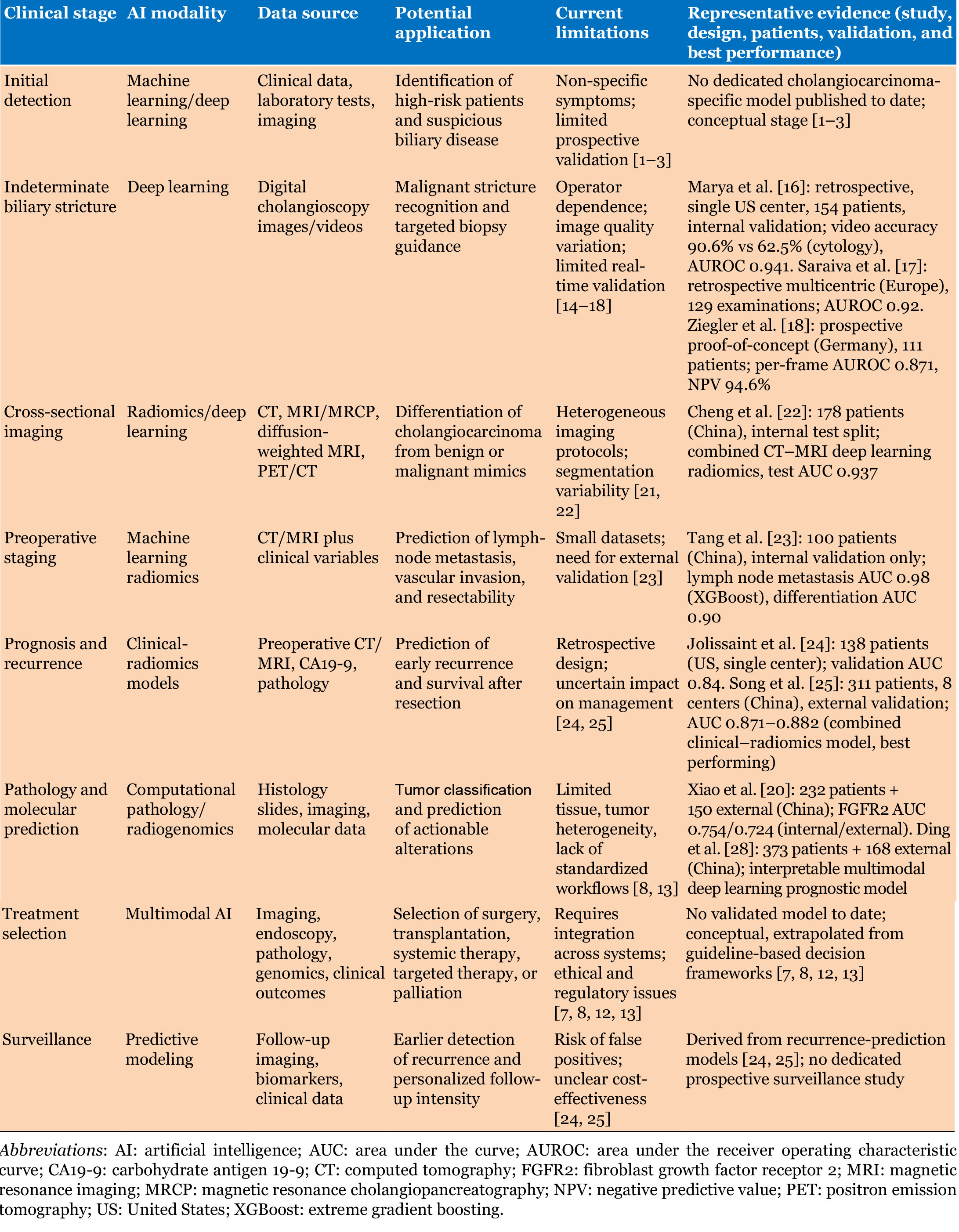
Each of these challenges can be mapped directly onto a candidate AI application, and this mapping provides the structure for the sections that follow. The subjective, operator-dependent visual assessment of indeterminate strictures corresponds to deep learning-based computer-aided detection and diagnosis during cholangioscopy; false-negative brush cytology and forceps biopsy correspond to AI-guided biopsy targeting; the difficulty of radiological differentiation between benign and malignant disease corresponds to radiomics-based classification of CT and MRI/MRCP; uncertainty regarding staging, nodal involvement, and resectability corresponds to machine learning prediction models built on preoperative imaging; limited tissue availability for molecular profiling corresponds to computational pathology tools that triage specimens for genomic testing; and the fragmentation of multimodal information at the tumor board corresponds to integrated clinical decision-support models (Table 1).
AI IN INDETERMINATE BILIARY STRICTURES AND CHOLANGIOSCOPY
Indeterminate biliary strictures are one of the most clinically relevant entry points for artificial intelligence in cholangiocarcinoma care. Diagnostic uncertainty arises because malignant and benign strictures may share overlapping cholangiographic, endoscopic, and radiological appearances, and conventional tissue acquisition may be falsely negative. Digital single-operator cholangioscopy has improved the direct visualization of the biliary mucosa and allows targeted biopsy; however, interpretation remains operator-dependent. Visual features such as irregular mucosa, abnormal tumor vessels, papillary projections, ulceration, nodularity, and friability may suggest malignancy; however, these findings are subjective and difficult to standardize across centers and levels of expertise [14],[15].
Therefore, deep learning has been explored as a method for standardizing the visual diagnosis during cholangioscopy. Convolutional neural networks (CNNs) can be trained on large datasets of cholangioscopy images or videos to classify strictures as benign or malignant and recognize suspicious mucosal features. In a study from the United States, a cholangioscopy-based deep learning system was developed using 2,388,439 still images from 154 patients with biliary strictures. In video analysis, the model identified patients with malignant strictures with an accuracy of 90.6%, compared with 62.5% for brush cytology (P = .04) and 60.9% for forceps biopsy (P = .03), and achieved an area under the receiver operating characteristic curve (AUROC) of 0.941 for the classification of high-quality malignant images [16]. This is clinically important because AI may provide real-time visual support for endoscopic decision-making, particularly when tissue acquisition is difficult, sampling is limited, or the endoscopist is uncertain whether further targeted biopsies are required [16].
More recent multicenter studies have expanded this concept by using digital cholangioscopy images to develop deep learning models for the automatic diagnosis and morphological characterization of malignant biliary strictures. In a European multicentric study coordinated in Porto, Portugal, a CNN trained on 84,994 images from 129 digital cholangioscopy examinations achieved an overall accuracy of 82.9%, a sensitivity of 83.5%, a specificity of 82.4%, and an AUROC of 0.92 for the automatic detection and morphological characterization of malignant strictures [17]. These systems aim not only to classify strictures as malignant or benign, but also to identify visual patterns associated with malignant transformation. In principle, such models can support computer-aided detection by highlighting suspicious areas, and computer-aided diagnosis by estimating the probability of malignancy. However, most available studies are retrospective or proof-of-concept, and the performance of these algorithms may be affected by image quality, bile, blood, debris, inflammation, stents, lighting, device generation, and differences in acquisition protocols [17].
AI may also enhance broader endoscopic evaluation of suspected cholangiocarcinoma by integrating cholangioscopy with clinical, biochemical, cytological, and molecular data. In a German proof-of-concept study developed through an academic–industry collaboration, a multimodal CNN for the real-time detection and differentiation of malignant and inflammatory strictures was trained on 15,158 cholangioscopy frames from 111 patients; on a test set of 5,715 frames from 20 patients, it achieved a per-frame AUROC of 0.871, a sensitivity of 80.9%, a specificity of 77.3%, a positive predictive value of 45.0%, and a negative predictive value of 94.6% [18]. A purely image-based model may misclassify inflammatory or PSC-related strictures as malignant, whereas a multimodal model can incorporate age, PSC status, bilirubin level, carbohydrate antigen 19-9 (CA19-9) level, dominant stricture characteristics, cytology, fluorescence in situ hybridization, and cross-sectional imaging findings. This approach is attractive because the clinical diagnosis of cholangiocarcinoma is rarely based on a single test. Instead, malignancy risk is inferred from multiple imperfect signals, making indeterminate biliary strictures particularly suitable for AI-supported probabilistic decision making [18].
Summarized performance data from these studies suggest that AI-assisted cholangioscopy can reach, and in image-based analyses exceed, the diagnostic performance of conventional sampling. In a systematic review of artificial intelligence in endoscopic imaging for malignant biliary strictures, CNN-based cholangioscopy models (934 patients; 3,775,819 images) achieved an average accuracy of 94.9%, sensitivity of 94.7%, and specificity of 92.1%, with image processing times of 7–15 milliseconds per frame, supporting the technical feasibility of real-time use [14]. Nevertheless, these headline metrics should be interpreted cautiously because they are derived predominantly from retrospective, internally validated datasets with enriched disease prevalence; the substantially lower positive predictive value observed in prospective real-time testing illustrates how performance can fall when models leave curated development conditions [18].
Despite this promise, AI-assisted cholangioscopy should be viewed as an adjunct, rather than a replacement, for expert assessment and tissue diagnosis. In particular, the precise clinical role of these tools should not be overstated: on the basis of the current evidence, they function primarily as aids to lesion recognition and to biopsy targeting or biopsy assessment, rather than as independent diagnostic tools. No AI system has regulatory approval for the autonomous diagnosis of biliary strictures, and a management-defining diagnosis of malignancy should not rest on AI output alone; histological confirmation remains the reference standard. Important unanswered questions include whether AI improves the diagnostic yield in prospective clinical practice, reduces unnecessary surgery or repeated ERCP, improves targeted biopsy selection, and performs reliably in high-risk populations, such as patients with PSC. Future systems require external validation, real-time testing, transparent performance reporting, and integration into endoscopic workflows before they can be adopted in routine care. Currently, the most realistic near-term role of AI is to support endoscopists by improving the recognition of suspicious mucosal patterns, standardizing reporting, and helping to prioritize areas for biopsy during cholangioscopy [14],[18].
AI-assisted biopsy targeting and assessment may also contribute directly to histopathological diagnosis through several complementary routes. First, by highlighting the mucosal areas with the highest probability of malignancy in real time, computer-aided detection may help endoscopists select optimal biopsy sites, potentially improving tissue sampling adequacy and reducing the sampling error that is a principal cause of false-negative results in desmoplastic and infiltrative tumors [14],[15]. Second, computational pathology can assist pathologists in evaluating the biopsy material itself: in perihilar cholangiocarcinoma, deep learning models applied to digitized biopsy and resection slides have been explored for tumor detection as a first step toward AI-supported histopathological assessment [19]. Third, image analysis may extend beyond diagnosis to biomarker prediction, as histopathology-based systems can screen routine hematoxylin and eosin sections for actionable genetic alterations [20]. Finally, models that integrate endoscopic imaging, radiological findings, and histopathological and molecular data are the most plausible route by which AI could shorten the diagnostic delay associated with repeated, inconclusive sampling [15].
AI AND RADIOMICS IN CROSS-SECTIONAL IMAGING
Cross-sectional imaging is central to the diagnosis, staging, and treatment planning of cholangiocarcinoma, making it one of the most important domains for AI applications. Computed tomography, MRI/MRCP, diffusion-weighted MRI, and positron emission tomography (PET)/CT provide complementary information on the tumor location, biliary involvement, vascular invasion, nodal disease, metastatic spread, and postoperative recurrence risk. However, conventional imaging interpretation remains partly subjective and may be affected by the tumor subtype, inflammatory changes, biliary obstruction, prior stenting, imaging protocol variation, and radiologist’s experience. Radiomics attempts to address some of these limitations by extracting quantitative imaging features related to tumor shape, intensity, texture, heterogeneity, and spatial relationships, which can then be analyzed using machine-learning or deep-learning models [21].
AI-based imaging models for intrahepatic cholangiocarcinoma have been investigated for lesion characterization and differential diagnosis. This is clinically relevant because intrahepatic cholangiocarcinoma can mimic hepatocellular carcinoma, combined hepatocellular-cholangiocarcinoma, and metastatic adenocarcinoma, particularly when imaging findings are atypical. Deep-learning radiomics models using CT and MRI have shown potential for improving the classification of intrahepatic cholangiocarcinoma among primary liver cancers, and MRI-based radiomics has also been explored to distinguish mass-forming intrahepatic cholangiocarcinoma from colorectal liver metastases. In a Chinese study of 178 patients (124 in the training and 54 in the test cohort), MRI-based deep learning radiomics outperformed the corresponding CT-based models for diagnosing intrahepatic cholangiocarcinoma [test-cohort area under the curve (AUC) 0.923 versus 0.880 for the best-performing models], and a combined CT–MRI cross-modal model reached an AUC of 0.937 in the test cohort [22]. These applications may be particularly valuable when tissue diagnosis is difficult, biopsy is inconclusive, or the imaging phenotype influences eligibility for surgery, locoregional therapy, or systemic treatment [22].
Radiomics has also been evaluated for preoperative risk stratification of extrahepatic and perihilar cholangiocarcinomas. Magnetic resonance imaging-based machine learning radiomics models have been developed to predict pathological differentiation and lymph node metastasis in extrahepatic cholangiocarcinoma, whereas CT-based radiomics approaches have been studied to predict nodal disease in perihilar cholangiocarcinoma. In a representative study of 100 patients with extrahepatic cholangiocarcinoma, machine learning radiomics models predicted lymph node metastasis with an AUC of 0.98 (accuracy 90%, sensitivity 75%, specificity 94%) and the degree of pathological differentiation with an AUC of 0.90 (accuracy 85%), although validation was internal only [23]. These predictions are clinically important, because lymph node metastasis, poor differentiation, and locally advanced disease strongly influence surgical planning, prognosis, and decisions regarding neoadjuvant or systemic therapy. If externally validated, AI-based imaging biomarkers can support multidisciplinary teams by identifying patients who require more intensive staging, altered operative strategies, or early systemic treatment consideration [23].
Another major area of interest is the prediction of recurrence after curative intent resection. Recurrence is common after surgery for intrahepatic cholangiocarcinoma, and conventional clinicopathological factors do not fully capture the biological aggressiveness before treatment. Machine learning radiomic models using preoperative CT have demonstrated the ability to predict early or very early recurrence after resection. At a single US cancer center, a model combining radiomic features with tumor size predicted early liver recurrence with an AUC of 0.84 [95% confidence interval (CI) 0.73–0.95] in the validation cohort of a 138-patient series [24]. In a Chinese study conducted across eight centers (311 patients), a combined clinical–radiomics model predicted early recurrence with an AUC of 0.974 in the derivation cohort and 0.871–0.882 in the internal and external validation cohorts, outperforming conventional American Joint Committee on Cancer (AJCC) tumor–node–metastasis (TNM) staging (AUC 0.686–0.717) [25]. Multicenter studies have suggested that combined clinical-radiomics models may outperform clinical models alone by integrating imaging heterogeneity with factors such as tumor stage, CA19-9, and histological features. These findings support the concept that imaging contains hidden information about tumor biology, which may help identify patients at high risk for early relapse [24],[25].
Despite these promising results, radiomics in cholangiocarcinoma are limited by methodological and clinical barriers. Many studies have been retrospective, single-center studies based on small or imbalanced datasets. Feature extraction can be affected by the segmentation technique, scanner type, contrast phase, reconstruction parameters, MRI sequence selection, and timing relative to biliary drainage or intervention. In addition, many models lack external validation, prospective testing, calibration analysis, interpretability, or evidence that their use improves patient outcomes. Therefore, while AI-based radiomics may become an important decision-support tool for cholangiocarcinoma, its current role is best viewed as investigational and complementary to expert radiological reviews, histopathology, molecular testing, and multidisciplinary discussions [21],[24]. The potential applications of AI across the cholangiocarcinoma care pathway, including endoscopic assessment, imaging interpretation, staging, molecular prediction, treatment selection, and surveillance, are summarized in Table 1.
AI IN MOLECULAR PREDICTION, PROGNOSIS, AND PERSONALIZED TREATMENT SELECTION
The role of artificial intelligence in cholangiocarcinoma extends beyond image-based diagnosis to include molecular prediction, prognostic modeling, recurrence risk assessment, and treatment selection. This is particularly relevant because cholangiocarcinoma is biologically heterogeneous and its management increasingly depends on integrating the anatomical subtype, tumor burden, histology, molecular profile, liver function, performance status, and expected treatment response. In this setting, AI may help transform fragmented diagnostic information into clinically meaningful risk stratification, supporting more individualized decisions across surgery, systemic therapy, targeted therapy, immunotherapy, locoregional treatment, biliary drainage, and surveillance [20].
Computational pathology is an emerging field in which AI may assist in risk stratification and the prediction of biomarkers. Whole-slide imaging allows digital analysis of tumor architecture, stromal composition, glandular morphology, necrosis, immune cell distribution, and spatial heterogeneity. In intrahepatic cholangiocarcinoma, a histopathology-based AI system developed at a Chinese academic center and trained on 2,069 whole-slide images from 232 patients predicted FGFR2 alterations with an AUC of 0.754 (0.724 in an external cohort of 150 patients) and IDH alterations with an AUC of 0.713 (0.656 externally) from routine hematoxylin and eosin slides, suggesting that morphological patterns correlate with the underlying molecular features; in the same study, modeled cost per progression-free quality-adjusted life month was substantially lower with AI-assisted triage than with a sequence-all strategy ($13,871.72 versus $44,538.93) [20]. This approach is not intended to replace molecular testing but may help prioritize patients for comprehensive genomic profiling, especially when tissue availability, cost, or turnaround time are limiting factors [19],[20].
Molecular profiling has become increasingly important in advanced cholangiocarcinoma, because actionable alterations may guide treatment. Relevant targets include FGFR2 fusions or rearrangements, IDH1 mutations,
HER2 alterations, BRAF mutations, NTRK fusions, and microsatellite instability or mismatch repair deficiency. AI-based pathology and radiogenomic models may support earlier identification of patients likely to harbor such alterations, allowing for more efficient use of confirmatory molecular testing and earlier consideration of targeted treatments. Computed tomography-based radiogenomic approaches have also been explored to predict the molecular status of intrahepatic cholangiocarcinoma, reinforcing the concept that imaging phenotypes may reflect tumor biology [26],[27].
Artificial intelligence may also contribute to prognostic modeling and the prediction of recurrence. Recurrence remains common after curative-intent resection, and conventional clinicopathological factors do not always fully capture tumor aggressiveness before treatment. Clinical radiomics and machine learning models may improve the prediction of early recurrence, survival, lymph node metastasis, vascular invasion, and postoperative outcomes by combining imaging features with biochemical, pathological, and clinical data. These tools can help identify patients who may benefit from intensified staging, closer surveillance, adjuvant treatment, neoadjuvant strategies, or early systemic therapy. However, most models are investigational and require prospective validation before they can be used to alter treatment pathways [24],[25],[28].
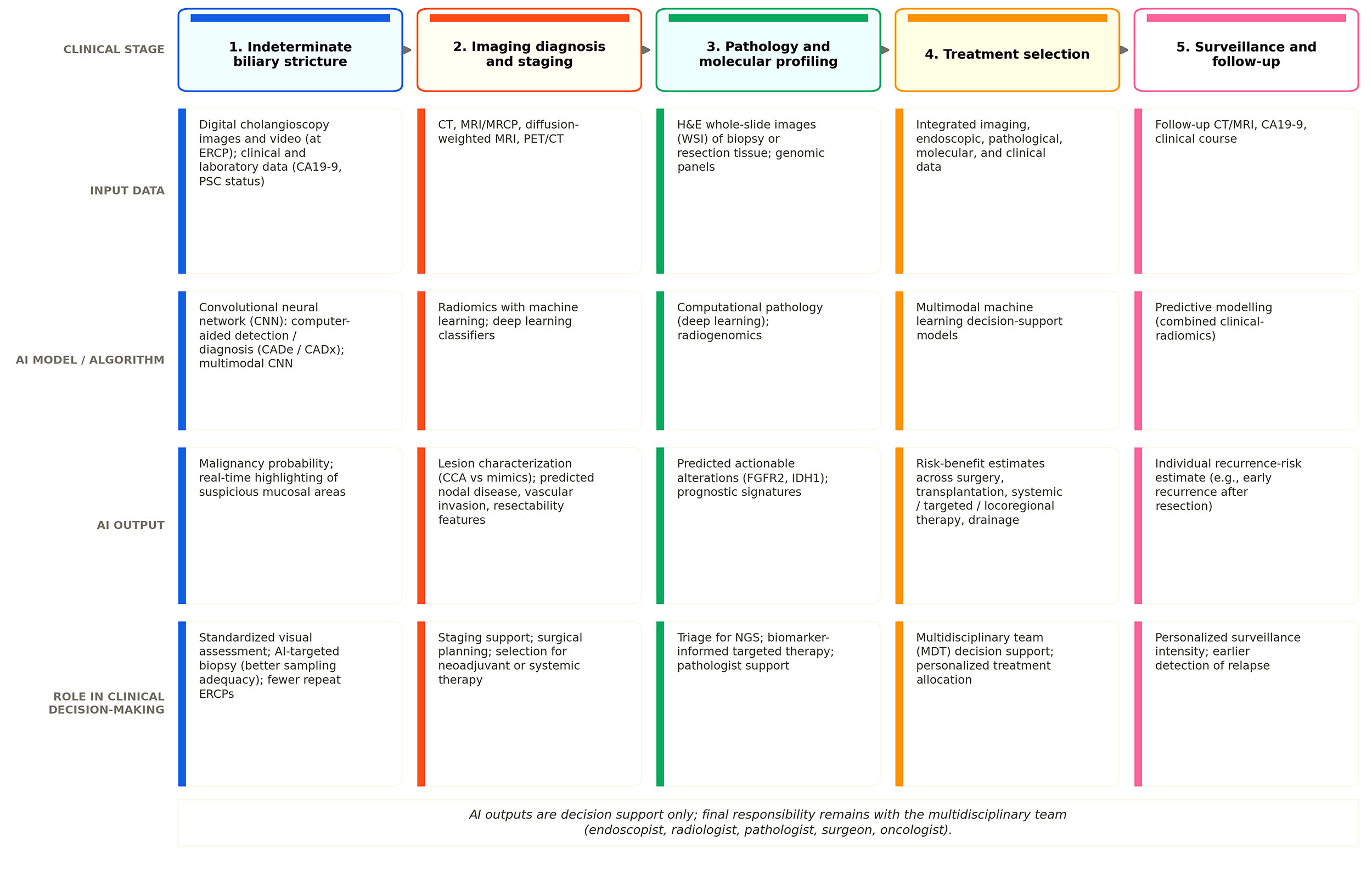
The most clinically meaningful future application of AI is personalized treatment selection. In localized diseases, AI-supported models can help estimate the resectability, future liver remnant risk, lymph node involvement, and recurrence probability. In selected perihilar cholangiocarcinomas, integrated models may help assess the suitability of transplant-based protocols, although such decisions must remain highly specialized and multidisciplinary. In advanced disease, AI can potentially combine molecular alterations, tumor burden, imaging phenotype, laboratory values, performance status, and prior treatment response to support selection among chemotherapy, immunotherapy, targeted therapy, locoregional therapy, endoscopic or percutaneous drainage, and the best supportive care. The proposed framework for integrating AI across the cholangiocarcinoma pathway, from diagnostic evaluation to molecular prediction, risk stratification, treatment selection, and surveillance, is shown in Figure 1 [7],[8],[12],[13],[29].
At present, these applications should be viewed as decision-support tools rather than as autonomous decision-makers. Cholangiocarcinoma management requires nuanced clinical judgment, particularly when evidence is limited, the tissue is inadequate, biliary sepsis is present, or the patient’s physiological reserve changes rapidly. The practical value of AI depends on whether the models can provide interpretable, externally validated, and clinically actionable outputs that improve the decisions made by multidisciplinary teams. The ultimate aim is not to replace expert clinicians, radiologists, endoscopists, pathologists, surgeons, and oncologists but to support earlier diagnosis, better risk stratification, and more precise treatment allocation [28],[29].
AI IN CHOLANGIOCARCINOMA COMPARED WITH RADIOLOGY, PATHOLOGY, AND OTHER SPECIALTIES
Placing cholangiocarcinoma-focused AI in the context of neighboring specialties clarifies both its promise and its relative immaturity. In radiology, AI-assisted image analysis has progressed considerably further along the translational pathway: by 2020, 100 Conformité Européenne (CE)-marked AI products from 54 vendors were already commercially available for clinical radiology in Europe. Even in this comparatively mature market, however, 64 of 100 products had no peer-reviewed evidence of efficacy, and only 18 of 100 had evidence at the level of demonstrated (potential) clinical impact, illustrating that regulatory clearance does not by itself guarantee robust clinical validation [30].
In pathology, clinical-grade AI has reached routine diagnostic deployment in selected use cases. An automated prostate cancer detection system (Paige Prostate, the first AI-based pathology tool authorized by the United States Food and Drug Administration) achieved a sensitivity of 0.99 and a negative predictive value of 1.0 in an independent, real-world validation performed in Brazil, and reduced diagnostic time by 65.5%, demonstrating what mature, externally validated computational pathology can deliver [31]. In gastrointestinal endoscopy, computer-aided polyp detection during colonoscopy is supported by randomized controlled trials: a meta-analysis of five trials including 4,354 patients showed a pooled adenoma detection rate of 36.6% with computer-aided detection versus 25.2% with conventional colonoscopy (relative risk 1.44, 95% CI 1.27–1.62), and such systems have received regulatory clearance and entered routine practice in several regions [32].
By comparison, AI for cholangiocarcinoma remains at the research stage: no AI device has been approved specifically for biliary stricture diagnosis or cholangiocarcinoma management, development cohorts are one to two orders of magnitude smaller, and randomized evidence is absent. This gap is explained partly by the rarity of the disease, the heterogeneity of its anatomical subtypes, the difficulty of establishing ground truth in indeterminate strictures, and the invasive, operator-dependent nature of cholangioscopy image acquisition. Nevertheless, the trajectory observed in radiology, pathology, and colonoscopy (from retrospective accuracy studies, through multicenter external validation, to prospective and randomized evaluation with post-market surveillance) offers a realistic translational template for the biliary field [14],[30],[32].
To date, the systems closest to clinical translation in cholangiocarcinoma are research models developed at academic centers rather than commercial platforms: cholangioscopy-based CNNs developed in the United States [16], a multicenter European deep learning model developed in Portugal and Spain [17], a real-time multimodal system developed in Germany through an academic–industry collaboration [18], multicenter radiomics models for recurrence prediction developed in China [25], and computational pathology systems for biomarker screening and prognostication developed at Chinese academic hospitals [20],[28]. Their geographic distribution, concentrated in the United States, Western Europe, and China, mirrors the availability of digital cholangioscopy, imaging infrastructure, and annotated datasets, and highlights the current underrepresentation of high-incidence, resource-limited regions in AI development.
GUIDELINES, REGULATION, AND GOVERNANCE OF CLINICAL AI
The translation of these tools into practice is increasingly shaped by formal guidance covering the development, validation, reporting, and clinical use of medical AI. Reporting frameworks now exist for every stage of the AI life cycle: the Transparent Reporting of a multivariable prediction model for Individual Prognosis Or Diagnosis (TRIPOD)+AI statement for diagnostic and prognostic prediction models [33], the Checklist for Artificial Intelligence in Medical Imaging (CLAIM) for imaging studies [34], the Developmental and Exploratory Clinical Investigations of DEcision support systems driven by Artificial Intelligence (DECIDE-AI) guideline for the early, live clinical evaluation of AI-based decision support [35], and the Standard Protocol Items: Recommendations for Interventional Trials (SPIRIT)-AI and Consolidated Standards of Reporting Trials (CONSORT)-AI extensions for the protocols and reports of clinical trials of AI interventions [36],[37]. Adherence to these frameworks directly addresses many of the weaknesses of the cholangiocarcinoma literature summarized in this review, including incomplete descriptions of patient selection, image preprocessing, validation strategy, and the handling of missing data.
Beyond reporting, the World Health Organization has articulated six governance principles for health AI: protecting human autonomy; promoting human well-being, safety, and the public interest; ensuring transparency and explainability; fostering responsibility and accountability; ensuring inclusiveness and equity; and promoting responsive and sustainable systems [38]. Regulatory agencies increasingly treat clinical AI as software as a medical device, with pre-market review and post-market surveillance requirements in the United States and the European Union, where high-risk health applications additionally fall within the scope of the European Union Artificial Intelligence Act.
These considerations are particularly consequential in high-burden and resource-constrained settings. In the Indian healthcare setting, for example, the Indian Council of Medical Research has published dedicated ethical guidelines for the application of AI in biomedical research and healthcare, emphasizing patient safety, informed consent, data privacy, transparency, accountability, and continuing clinical oversight of AI outputs [39]; software intended for medical purposes is regulated as a medical device by the national regulator, and the Digital Personal Data Protection Act (2023) governs the processing of personal health data. Comparable quality-assurance expectations (local validation before deployment, clear allocation of responsibility between clinician and tool, mechanisms for auditing performance drift, and protection of the patient data used for model training) will apply wherever AI tools for cholangiocarcinoma are eventually implemented, and they are especially important in regions where the disease burden is highest but digital infrastructure and regulatory capacity may be limited.
LIMITATIONS, IMPLEMENTATION CHALLENGES, AND FUTURE DIRECTIONS
Despite significant advancements, the majority of artificial intelligence applications in cholangiocarcinoma remain at an early developmental stage. Many existing studies are retrospective, limited to single centers, and rely on relatively small datasets due to the disease’s rarity and diversity. Cholangiocarcinomas encompass intrahepatic, perihilar, and distal subtypes, each characterized by distinct imaging features, biological behaviors, treatment protocols, and prognoses. While aggregating these categories may enhance sample size for analysis purposes, it poses a risk of diminishing clinical specificity; conversely, examining them individually frequently results in models that lack sufficient power. Class imbalances, limited annotation quality, and inconsistent reference standards restrict the model reliability. These issues are particularly important in indeterminate biliary strictures, where false-negative tissue samples and overlapping benign inflammatory appearances may complicate the definition of the ground truth [35].
A critical appraisal of the underlying evidence reinforces this caution. Most published models were developed retrospectively, and truly external, geographically independent validation remains the exception rather than the rule. Among the cholangioscopy studies, the US deep learning model was trained and tested on data from a single high-volume center (154 patients) without external validation [16]; the European multicentric model was retrospective and validated on an internal image split (129 examinations) [17]; and the German real-time system, although evaluated prospectively, was a proof-of- concept study of 111 patients with a test set drawn from only 20 patients [18]. Representative radiomics studies show the same pattern of small, often imbalanced cohorts (100–178 patients in the diagnostic and staging studies discussed above) with internal validation only [22],[23],[24]. Where external validation has been performed, performance typically falls: the eight-center recurrence model declined from an AUC of 0.974 in derivation to 0.871–0.882 in validation cohorts [25], and the histopathology-based biomarker system declined from an AUC of 0.754 to 0.724 for FGFR2 and from 0.713 to 0.656 for IDH on external testing, a predictable consequence of overfitting and cohort selection [20]. Selection bias is compounded by the tertiary-center origin of most cohorts, disease prevalence enriched well above that of routine practice, the exclusion of low-quality images and incomplete records, and heterogeneous reference standards. Generalizability to community settings, other populations, and different device generations is therefore unproven, and no model has yet been shown in a prospective comparative study to change management or improve patient-relevant outcomes.
Technical barriers limit reproducibility and clinical translation. Radiomics models are sensitive to scanner type, contrast phase, reconstruction parameters, MRI sequences, segmentation methods, and timing relative to biliary drainage or intervention. Cholangioscopy-based models are affected by bile, blood, debris, inflammation, stents, lighting conditions, and device generation. Computational pathology models require standardized slide preparation, digitization, annotation, and quality control. In addition, many AI systems have been developed on highly selected datasets and may perform poorly when applied to external populations, different centers, or real-world workflows. Transparent reporting, external validation, calibration, and prospective testing are essential before these models are considered clinically dependable [35],[36],[37].
Implementation requires more than diagnostic accuracy. AI tools must provide outputs that are interpretable, timely, and actionable for multidisciplinary decision making. A model that predicts malignancy, recurrence, or molecular status is useful only if it changes management strategies to improve outcomes; reduce unnecessary procedures; accelerate appropriate treatment; or improve patient selection for surgery, transplantation, systemic therapy, targeted therapy, or surveillance. Important practical questions remain regarding medicolegal responsibility, clinician trust, data governance, cost-effectiveness, equity, regulatory approval, and integration into endoscopy, radiology, pathology, oncology, and tumor board workflows. Overreliance on AI must also be avoided, particularly in complex cases where the clinical context, patient preference, tissue adequacy, liver function, biliary sepsis, and treatment fitness remain decisive [34],[37].
Future research should focus on prospective, multicenter, externally validated studies that evaluate AI tools in a clinically realistic setting. Priority areas include real-time AI-assisted cholangioscopy for indeterminate biliary strictures; standardized radiomics pipelines for CT and MRI/MRCP; computational pathology tools linked to molecular testing; and multimodal models combining imaging, endoscopy, histology, genomics, laboratory data, and treatment outcomes. Instead of developing isolated algorithms, future systems should be designed as decision-support platforms that complement multidisciplinary expertise. If validated responsibly, AI may help move cholangiocarcinoma care toward earlier diagnosis, better risk stratification, biomarker-informed therapy, individualized surveillance, and more consistent treatment selection across institutions [34],[36].
Conclusion
Artificial intelligence has the potential to support care for multiple stages of cholangiocarcinoma, from the assessment of indeterminate biliary strictures to imaging interpretation, molecular prediction, prognostic modeling, treatment selection, and surveillance. Artificial intelligence-assisted cholangioscopy may help standardize visual assessment and guide targeted biopsy, whereas radiomics and radiogenomics may extract clinically relevant information from CT, MRI/MRCP, PET/CT, and histopathology that is not readily apparent through conventional interpretation. These applications are particularly relevant in cholangiocarcinoma, where delayed diagnosis, limited tissue yield, tumor heterogeneity, and complex multidisciplinary decision-making remain major clinical challenges.
However, AI should currently be regarded as an adjunct to expert clinical judgment, rather than a replacement for established diagnostic and therapeutic pathways. Most existing models necessitate external validation, prospective testing, transparent documentation, and incorporation into comprehensive multidisciplinary workflows before they can be routinely implemented. The future potential of AI in the context of cholangiocarcinoma is expected to hinge on multimodal systems that integrate endoscopic, radiological, pathological, molecular, and clinical information. This integration aims to facilitate earlier diagnoses, enhance risk stratification accuracy, inform treatment through biomarkers, and provide personalized follow-up care.
NOVELTY OF THE STUDY
To the best of the authors’ knowledge, this is among the first reviews to follow artificial intelligence across the entire cholangiocarcinoma pathway, from the initial evaluation of indeterminate biliary strictures, through cholangioscopy, cross-sectional imaging, computational pathology, and molecular prediction, to personalized treatment selection and surveillance, rather than addressing a single modality in isolation. The review explicitly pairs each major diagnostic and therapeutic challenge with the AI application designed to address it, summarizes the quantitative diagnostic performance and validation status of representative models together with the settings in which they were developed, and benchmarks the maturity of biliary AI against the more advanced experience of radiology, pathology, and gastrointestinal endoscopy. In addition, it integrates the emerging reporting, regulatory, and governance frameworks for clinical AI, including those relevant to high-burden and resource-limited settings, into a single clinically oriented synthesis. This combined clinical, technical, and governance perspective is intended to give practicing clinicians a realistic map of what AI can and cannot yet contribute to cholangiocarcinoma care, and to identify the validation steps required before routine adoption.
REFERENCES
1.
Ilyas SI, Affo S, Goyal L, Lamarca A, Sapisochin G, Yang JD, et al. Cholangiocarcinoma – Novel biological insights and therapeutic strategies. Nat Rev Clin Oncol 2023;20(7):470–86. [CrossRef]
[Pubmed]

2.
Khan SA, Tavolari S, Brandi G. Cholangiocarcinoma: Epidemiology and risk factors. Liver Int 2019;39 Suppl 1:19–31. [CrossRef]
[Pubmed]
3.
Banales JM, Marin JJG, Lamarca A, Rodrigues PM, Khan SA, Roberts LR, et al. Cholangiocarcinoma 2020: The next horizon in mechanisms and management. Nat Rev Gastroenterol Hepatol 2020;17(9):557–88. [CrossRef]
[Pubmed]
4.
Zerunian M, Polidori T, Palmeri F, Nardacci S, Del Gaudio A, Masci B, et al. Artificial intelligence and radiomics in cholangiocarcinoma: A comprehensive review. Diagnostics (Basel) 2025;15(2):148. [CrossRef]
[Pubmed]
5.
Kamsa-Ard S, Santong C, Kamsa-Ard S, Luvira V, Luvira V, Suwanrungruang K, et al. Decreasing trends in cholangiocarcinoma incidence and relative survival in Khon Kaen, Thailand: An updated, inclusive, population-based cancer registry analysis for 1989–2018. PLoS One 2021;16(2):e0246490. [CrossRef]
[Pubmed]
6.
Rushbrook SM, Kendall TJ, Zen Y, Albazaz R, Manoharan P, Pereira SP, et al. British Society of Gastroenterology Guidelines for the diagnosis and management of cholangiocarcinoma. Gut 2023;73(1):16–46. [CrossRef]
[Pubmed]
7.
Vogel A, Bridgewater J, Edeline J, Kelley RK, Klümpen HJ, Malka D, et al. Biliary tract cancer: ESMO Clinical Practice Guideline for diagnosis, treatment and follow-up. Ann Oncol 2023;34(2):127–40. [CrossRef]
[Pubmed]
8.
Vogel A, Ducreux M; ESMO Guidelines Committee. ESMO Clinical Practice Guideline interim update on the management of biliary tract cancer. ESMO Open 2025;10(1):104003. [CrossRef]
[Pubmed]
9.
Navaneethan U, Njei B, Venkatesh PG, Lourdusamy V, Sanaka MR. Endoscopic ultrasound in the diagnosis of cholangiocarcinoma as the etiology of biliary strictures: A systematic review and meta-analysis. Gastroenterol Rep (Oxf) 2015;3(3):209–15. [CrossRef]
[Pubmed]
10.
Fujii-Lau LL, Thosani NC, Al-Haddad M, Acoba J, Wray CJ, Zvavanjanja R, et al. American Society for Gastrointestinal Endoscopy Guideline on the role of endoscopy in the diagnosis of malignancy in biliary strictures of undetermined etiology: Summary and recommendations. Gastrointest Endosc 2023;98(5):685–93. [CrossRef]
[Pubmed]
11.
Razumilava N, Gores GJ. Cholangiocarcinoma. Lancet 2014;383(9935):2168–79. [CrossRef]
[Pubmed]
12.
Buckholz AP, Brown RS Jr. Cholangiocarcinoma: Diagnosis and management. Clin Liver Dis 2020;24(3):421–36. [CrossRef]
[Pubmed]
13.
Valle JW, Kelley RK, Nervi B, Oh DY, Zhu AX. Biliary tract cancer. Lancet 2021;397(10272):428–44. [CrossRef]
[Pubmed]
14.
Njei B, McCarty TR, Mohan BP, Fozo L, Navaneethan U. Artificial intelligence in endoscopic imaging for detection of malignant biliary strictures and cholangiocarcinoma: A systematic review. Ann Gastroenterol 2023;36(2):223–30. [CrossRef]
[Pubmed]
15.
Tang RSY. Endoscopic evaluation of indeterminate biliary strictures: Cholangioscopy, endoscopic ultrasound, or both? Dig Endosc 2024;36(7):778–88. [CrossRef]
[Pubmed]
16.
Marya NB, Powers PD, Petersen BT, Law R, Storm A, Abusaleh RR, et al. Identification of patients with malignant biliary strictures using a cholangioscopy-based deep learning artificial intelligence (with video). Gastrointest Endosc 2023;97(2):268–78.e1. [CrossRef]
[Pubmed]
17.
Saraiva MM, Ribeiro T, González-Haba M, Agudo Castillo B, Ferreira JPS, Vilas Boas F, et al. Deep learning for automatic diagnosis and morphologic characterization of malignant biliary strictures using digital cholangioscopy: A multicentric study. Cancers (Basel) 2023;15(19):4827. [CrossRef]
[Pubmed]
18.
Ziegler J, Dobsch P, Rozema M, Zuber-Jerger I, Weigand K, Reuther S, et al. Multimodal convolutional neural network-based algorithm for real-time detection and differentiation of malignant and inflammatory biliary strictures in cholangioscopy: A proof-of-concept study (with video). Gastrointest Endosc 2025;101(4):830–42.e2. [CrossRef]
[Pubmed]
19.
Hoyer DP, Ting S, Rogacka N, Koitka S, Hosch R, Flaschel N, et al. AI-based digital histopathology for perihilar cholangiocarcinoma: A step, not a jump. J Pathol Inform 2023;15:100345. [CrossRef]
[Pubmed]
20.
Xiao H, Wang J, Weng Z, Lin X, Shu M, Shen J, et al. A histopathology-based artificial intelligence system assisting the screening of genetic alteration in intrahepatic cholangiocarcinoma. Br J Cancer 2025;132(2):195–202. [CrossRef]
[Pubmed]
21.
Brunese MC, Fantozzi MR, Fusco R, De Muzio F, Gabelloni M, Danti G, et al. Update on the applications of radiomics in diagnosis, staging, and recurrence of intrahepatic cholangiocarcinoma. Diagnostics (Basel) 2023;13(8):1488. [CrossRef]
[Pubmed]
22.
Cheng M, Zhang H, Guo Y, Lyu P, Yan J, Liu Y, et al. Comparison of MRI and CT based deep learning radiomics analyses and their combination for diagnosing intrahepatic cholangiocarcinoma. Sci Rep 2025;15(1):9629. [CrossRef]
[Pubmed]
23.
Tang Y, Yang CM, Su S, Wang WJ, Fan LP, Shu J. Machine learning-based radiomics analysis for differentiation degree and lymphatic node metastasis of extrahepatic cholangiocarcinoma. BMC Cancer 2021;21(1):1268. [CrossRef]
[Pubmed]
24.
Jolissaint JS, Wang T, Soares KC, Chou JF, Gönen M, Pak LM, et al. Machine learning radiomics can predict early liver recurrence after resection of intrahepatic cholangiocarcinoma. HPB (Oxford) 2022;24(8):1341–50. [CrossRef]
[Pubmed]
25.
Song Y, Zhou G, Zhou Y, Xu Y, Zhang J, Zhang K, et al. Artificial intelligence CT radiomics to predict early recurrence of intrahepatic cholangiocarcinoma: A multicenter study. Hepatol Int 2023;17(4):1016–27. [CrossRef]
[Pubmed]
26.
Normanno N, Martinelli E, Melisi D, Pinto C, Rimassa L, Santini D, et al. Role of molecular genetics in the clinical management of cholangiocarcinoma. ESMO Open 2022;7(3):100505. [CrossRef]
[Pubmed]
27.
Viganò L, Zanuso V, Fiz F, Cerri L, Laino ME, Ammirabile A, et al. CT-based radiogenomics of intrahepatic cholangiocarcinoma. Dig Liver Dis 2025;57(1):118–24. [CrossRef]
[Pubmed]
28.
Ding GY, Tan WM, Lin YP, Ling Y, Huang W, Zhang S, et al. Mining the interpretable prognostic features from pathological image of intrahepatic cholangiocarcinoma using multi-modal deep learning. BMC Med 2024;22(1):282. [CrossRef]
[Pubmed]
29.
Stenzinger A, Vogel A, Lehmann U, Lamarca A, Hofman P, Terracciano L, et al. Molecular profiling in cholangiocarcinoma: A practical guide to next-generation sequencing. Cancer Treat Rev 2024;122:102649. [CrossRef]
[Pubmed]
30.
van Leeuwen KG, Schalekamp S, Rutten MJCM, van Ginneken B, de Rooij M. Artificial intelligence in radiology: 100 commercially available products and their scientific evidence. Eur Radiol 2021;31(6):3797–804. [CrossRef]
[Pubmed]
31.
da Silva LM, Pereira EM, Salles PG, Godrich R, Ceballos R, Kunz JD, et al. Independent real-world application of a clinical-grade automated prostate cancer detection system. J Pathol 2021;254(2):147–58. [CrossRef]
[Pubmed]
32.
Hassan C, Spadaccini M, Iannone A, Maselli R, Jovani M, Chandrasekar VT, et al. Performance of artificial intelligence in colonoscopy for adenoma and polyp detection: A systematic review and meta-analysis. Gastrointest Endosc 2021;93(1):77–85.e6. [CrossRef]
[Pubmed]
33.
Collins GS, Moons KGM, Dhiman P, Riley RD, Beam AL, Van Calster B, et al. TRIPOD+AI statement: Updated guidance for reporting clinical prediction models that use regression or machine learning methods. BMJ 2024;385:e078378. [CrossRef]
[Pubmed]
34.
Tejani AS, Klontzas ME, Gatti AA, Mongan JT, Moy L, Park SH, et al. Checklist for Artificial Intelligence in Medical Imaging (CLAIM): 2024 update. Radiol Artif Intell 2024;6(4):e240300. [CrossRef]
[Pubmed]
35.
Vasey B, Nagendran M, Campbell B, Clifton DA, Collins GS, Denaxas S, et al. Reporting guideline for the early stage clinical evaluation of decision support systems driven by artificial intelligence: DECIDE-AI. BMJ 2022;377:e070904. [CrossRef]
[Pubmed]
36.
Cruz Rivera S, Liu X, Chan AW, Denniston AK, Calvert MJ; SPIRIT-AI and CONSORT-AI Working Group; SPIRIT-AI and CONSORT-AI Steering Group; SPIRIT-AI and CONSORT-AI Consensus Group. Guidelines for clinical trial protocols for interventions involving artificial intelligence: The SPIRIT-AI extension. Nat Med 2020;26(9):1351–63. [CrossRef]
[Pubmed]
37.
Liu X, Cruz Rivera S, Moher D, Calvert MJ, Denniston AK; SPIRIT-AI and CONSORT-AI Working Group. Reporting guidelines for clinical trial reports for interventions involving artificial intelligence: The CONSORT-AI extension. Nat Med 2020;26(9):1364–74. [CrossRef]
[Pubmed]
38.
World Health Organization. Ethics and governance of artificial intelligence for health: WHO guidance. Geneva: World Health Organization; 2021.
39.
Indian Council of Medical Research. Ethical guidelines for application of artificial intelligence in biomedical research and healthcare. New Delhi: Indian Council of Medical Research; 2023.
SUPPORTING INFORMATION
Acknowledgments
AI-generated content acknowledgement:
During the preparation of this manuscript, artificial intelligence-assisted language tools were used only to support language editing, grammar refinement, and improvement of readability. No AI tool was used to generate original scientific data, perform data analysis, create references, or replace the authors’ interpretation and critical judgment. The authors reviewed, edited, and approved all content and take full responsibility for the accuracy and integrity of the final manuscript.
Ahmed Salman - Conception of the work, Design of the work, Revising the work critically for important intellectual content, Final approval of the version to be published, Agree to be accountable for all aspects of the work in ensuring that questions related to the accuracy or integrity of any part of the work are appropriately investigated and resolved.
Ahmed Elewa - Conception of the work, Design of the work, Drafting the work, Final approval of the version to be published, Agree to be accountable for all aspects of the work in ensuring that questions related to the accuracy or integrity of any part of the work are appropriately investigated and resolved.
Ahmed Marwan - Conception of the work, Design of the work, Drafting the work, Final approval of the version to be published, Agree to be accountable for all aspects of the work in ensuring that questions related to the accuracy or integrity of any part of the work are appropriately investigated and resolved.
Guarantor of SubmissionThe corresponding author is the guarantor of submission.
Source of SupportNone
Data AvailabilityAll relevant data are within the paper and its Supporting Information files.
Conflict of InterestAuthors declare no conflict of interest.
Copyright© 2026 Ahmed Salman et al. This article is distributed under the terms of Creative Commons Attribution License which permits unrestricted use, distribution and reproduction in any medium provided the original author(s) and original publisher are properly credited. Please see the copyright policy on the journal website for more information.