

| Table of Contents |  |
|
Case Report
| ||||||
| Challenges in management of right aberrant hepatic duct injury | ||||||
| Jayashri S. Pandya1, Sudatta B. Waghmare2, Makrand M. Thakre3, Nilesh H. Doctor4 | ||||||
|
1M.S. General Surgery, Professor, General Surgery Department and Incharge Emergency Trauma services, T.N.M.C & B.Y.L. Nair Hospital, Mumbai, Maharashtra, India.
2M.S. General Surgery, Assistant Professor, General Surgery Department, T.N.M.C & B.Y.L. Nair Hospital, Mumbai, Maharashtra, India. 3PG Resident in General Surgery, Third year resident, General Surgery dept., T.N.M.C & B.Y.L. Nair Hospital, Mumbai, Maharashtra, India. 4M.S. General Surgery, Consultant Hepatobiliary Surgeon, Jaslok Hospital, Mumbai, Maharashtra, India. | ||||||
| ||||||
|
[HTML Abstract]
[PDF Full Text]
[Print This Article]
[Similar article in Pumed] [Similar article in Google Scholar] |


| How to cite this article: |
| Pandya JS, Waghmare SB, Thakre MM, Doctor NH. Challenges in management of right aberrant hepatic duct injury. Int J Hepatobiliary Pancreat Dis 2016;6:6–9. |
|
Abstract
|
|
Introduction:
Bile duct injury (BDI) is a known complication following laparoscopic cholecystectomy. Variation in biliary tree anatomy occurs in about 25% patients with aberrant right hepatic duct being the most common [1]. Low insertion of the right hepatic duct puts it at risk of damage during dissection of the Calot's triangle. The resulting isolated right posterior BDI poses a challenge for both diagnosis and management.
Case Report: We present a case of a persistent bile leak after laparoscopic cholecystectomy. Diagnosis of an aberrant right hepatic sectoral duct injury was confirmed by MRCP, and managed by Roux-en-Y hepaticojejunostomy. Conclusion: Right posterior hepatic duct opening located close to cystic duct is a dangerous scenario. Due to its location, it has high potential to get injured. The leak may then be erroneously attributed to injury to minor biliary radicle in gallbladder fossa and delay the diagnosis. Hence, there should be high index of suspicion to diagnose such injuries, as interpretation of ERCP images may be erroneous. | |
|
Keywords:
Bile duct injury, Cholecystectomy, Hepaticojejunostomy, Sectoral bile duct
| |
|
Introduction
| ||||||
|
Bile duct injury (BDI) is a serious complication of cholecystectomy. The incidence of aberrant biliary anatomy is approximately 1–1.5%, with anomalies of the right duct being the most frequent [2]. There are many factors which place the right posterior sectoral duct (RPSD) at risk of being ignored during cholecystectomy. These are: A low insertion of RPSD into the common bile duct close to the cystic duct, RPSD running behind the cystic duct to join the common bile duct, the cystic duct joining the confluence of the sectoral or hepatic ducts. | ||||||
|
Case Report
| ||||||
|
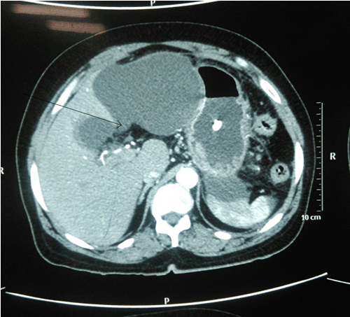
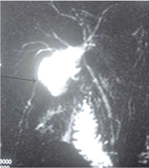
A 54-year-old female underwent uneventful laparoscopic cholecystectomy for acute cholecystitis. She developed pain in the right hypochondrium 4 days after surgery. There was no history of vomiting, fever or jaundice. Sonography showed a fluid collection in the gallbladder fossa, confirmed by computed tomography (CECT) scan (Figure 1). A 14-F Pigtail catheter was inserted to drain the collection. There was persistent drainage of 150–200 ml of bile per day. Endoscopic retrograde cholangiography (ERCP) was performed 10 days after initial surgery. It showed mildly dilated CBD without any obvious leak. The diagnosis of an isolated Strasberg type C bile duct injury [3] was made by magnetic resonance cholangiography (MRC) (Figure 3). On careful review of ERCP images, the right posterior hepatic duct was not visualized (Figure 2). The diagnosis of isolated Strasberg type C bile duct injury was confirmed. After period of six weeks the drainage remained 150-200 ml/day. A Roux-en-Y hepaticojejunostomy to the right aberrant hepatic duct was performed. Postoperative period was uneventful, liver function tests were normal. | ||||||
| ||||||
| ||||||
| ||||||
|
Discussion
| ||||||
|
Bile duct injury (BDI) is a recognized complication of laparoscopic cholecystectomy. Anomalous drainage of the right posterior sectoral bile duct into the cystic duct or the common hepatic duct is seen in around 2–5% of patients [1] increasing the risk of injury. Different BDI classifications are described in literature. Strasberg's classification [2] includes all types of injury including a leak. Right posterior sectoral bile duct injuries according to Strasberg's classification can be types B or C injuries. The operative injury to these ducts as a result of division or clipping can result in bile leak or recurrent episodes of cholangitis [4]. These patients usually present with abdominal pain or fever if biloma becomes infected. In some patients, injuries to these ducts may go unrecognized as the injured area tends to atrophy. Diagnosis and management protocol can be elusive. Abdominal ultrasound or CT-scan may reveal perihepatic fluid collection. Preoperative cholangiography is essential in defining the anatomy for a successful reconstruction following major bile duct injuries. The advantage of magnetic resonance cholangiopancreatography (MRCP) imaging is to delineate all ducts, even those that are excluded from the biliary system. The ERCP may be diagnostic and therapeutic. But ERCP has a limited role only in injuries where the connection of the extrahepatic tree to the duodenum is still intact. Bile leak in the presence of an intact common bile duct should raise the suspicion of RPSD injury [5] [6]. Intervention depends on the time period between surgery and recognition of the type, extent and level of the injury. External biliary drainage allows control of the leak and may obviate the need for surgery in 50% of patients [7]. In cases of persistent drainage after 8 weeks, a hepaticojejunostomy should be constructed to a Roux-en-Y jejunal limb. | ||||||
|
Conclusion
| ||||||
|
Right posterior sectoral duct (RPSD) opening located close to cystic duct is dangerous scenario. Due to its location, it has high potential to get injured. The leak may then be erroneously attributed to injury to minor biliary radicle in gall bladder fossa and delay the diagnosis. Hence, high index of suspicion to diagnose such injuries, as interpretation of ERCP images may be erroneous. | ||||||
|
References
| ||||||
| ||||||


|
[HTML Abstract]
[PDF Full Text]
|
|
Author Contributions
Jayashri S. Pandya – Substantial contributions to conception and design, Acquisition of data, Analysis and interpretation of data, Drafting the article, Revising it critically for important intellectual content, Final approval of the version to be published Sudatta B. Waghmare – Analysis and interpretation of data, Revising it critically for important intellectual content, Final approval of the version to be published Makrand M. Thakre – Analysis and interpretation of data, Revising it critically for important intellectual content, Final approval of the version to be published Nilesh H. Doctor – Analysis and interpretation of data, Revising it critically for important intellectual content, Final approval of the version to be published |
|
Guarantor of submission
The corresponding author is the guarantor of submission. |
|
Source of support
None |
|
Conflict of interest
Authors declare no conflict of interest. |
|
Copyright
© 2016 Jayashri S. Pandya et al. This article is distributed under the terms of Creative Commons Attribution License which permits unrestricted use, distribution and reproduction in any medium provided the original author(s) and original publisher are properly credited. Please see the copyright policy on the journal website for more information. |
|
|