| Table of Contents |  |
|
Original Article
|
| Clinicopathological significance of fatty acid synthase expression in pancreatic ductal carcinoma |
| Hiroshi Maekawa1, Tomoaki Ito2, Hajime Orita1, Koichi Sato3, Ryo Wada4 |
|
1Associate professor, department of surgery, Juntendo university school of medicine Shizuoka hospital, Izu-no-kuni city, Shizuoka, Japan.
2Assistant professor, department of surgery, Juntendo university school of medicine Shizuoka hospital, Izu-no-kuni city, Shizuoka, Japan. 3Professor, department of surgery, Juntendo university school of medicine Shizuoka hospital, Izu-no-kuni city, Shizuoka, Japan. 4Professor, department of pathology, Juntendo university school of medicine Shizuoka hospital, Izu-no-kuni city, Shizuoka, Japan. |
|
Article ID: 100011IJHPDHM2013 doi:10.5348/ijhpd-2013-11-OA-2 |
|
Address correspondence to: Hiroshi Maekawa 1129 Nagaoka, Izu-no-kuni Shizuoka Japan 410-2295 Phone: 81 55.948.3111 Fax: 81 55.946.0514 Email: hmaekawa0201@gmail.com |
|
[HTML Abstract]
[PDF Full Text]
|
| How to cite this article: |
| Maekawa H, Ito T, Orita H, Sato K, Wada R. Clinicopathological significance of fatty acid synthase expression in pancreatic ductal carcinoma. International Journal of Hepatobiliary and Pancreatic Diseases 2013;3:10–16. |
|
Abstract
|
|
Introduction:
Fatty Acid Synthase (FAS) expression is increased in various cancers and it contributes to tumor cellular activities. In clinical practice, pancreatic ductal carcinoma is regarded as a cancer with poor prognosis.
Aims: To investigated FAS expression in pancreatic ductal carcinoma (PDC) and compare with clinic-pathological features. Methods: We studied 38 patients who had resection for PDC in our hospital. We compared FAS staining with clinico-pathological features, including Ki-67 and p53 staining. Results: Using our criteria, out of 38 cases, 15 cases were considered FAS positive. FAS positivity correlated with Ki-67 positivity (p = 0.001), but there was no significant relationship between FAS expression and p53 staining (p = 0.224). FAS positive cases showed higher rate of lymph node metastases (p = 0.0038). FAS expression was related to differentiation of adenocarcinoma (p = 0.0347). Clinically, overall survival rate of FAS positive cases was lower than that of FAS negative cases (p = 0.0068). Conclusions: FAS expression may be related to lymph node metastases and clinical behavior in pancreatic ductal carcinoma. | |
|
Key Words:
Pancreatic cancer, Fatty acid synthase, Ki-67, Immunohistochemistry, Prognosis
| |
|
Introduction
| ||||||
|
In human tissues, fatty acid synthase (FAS) is usually expressed in hormone-sensitive and high lipid metabolism cells. [1] In normal tissues, FAS expression is regulated and inhibited by several hormones. [2] FAS expression is also increased in various cancers. Breast cancer or prostate cancer is a good example of cancers expressing FAS. [3] [4] [5] [6] [7] FAS expression increases de novo biosynthesis of fatty acids which contributes to tumor cellular activities; [8] metabolism for energy, building cellular membranes and intracellular second messengers. [9] [10] It has been demonstrated that cancer cells often express FAS and the intensity of FAS expression correlates with clinical outcome. Cases of high FAS expression often show poor prognosis in various types of cancers. [11] [12] [13] [14] [15] [16] [17] In clinical practice, pancreatic ductal carcinoma (PDC) is regarded as a cancer with poor prognosis. To investigate the clinicopathological significance of FAS expression in pancreatic ductal carcinoma (PDC), we studied patients surgically treated for PDC in our hospital. The aims of this study were, i) to investigate the relationship between the expression of FAS and clinicopathological features using immunohistochemistry in PDC, and ii) to analyze whether FAS may be considered as a prognostic marker in PDC. | ||||||
|
Materials and Methods
| ||||||
|
We studied 38 patients resected for PDC in our hospital, between January 2002 and January 2011. Clinical data such as gender, age, recurrence, and prognoses were obtained from clinical records. Pathological diagnosis was performed by examining hematoxylin and eosin stained sections. We investigated expressions of FAS, Ki-67, and p53 with immunohistochemical stainings on tissue sections cut from formalin-fixed and paraffin-embedded carcinoma specimens. We then compared FAS staining with clinical information and pathological aspects, including Ki-67 and p53 staining. The study protocol conformed to the ethical guidelines of the World Medical Association Declaration of Helsinki, and was approved by the ethical committee in our hospital. Immunohistochemical procedure for FAS expression: We used 4 µm tissue sections cut from formalin-fixed and paraffin-embedded specimens. Sections were deparaffinized by treatment with xylene for three minutes (two times) and rehydrated by passage through 100% ethanol for 30 sec (three times). Antigen retrieval was performed by microwaving slides in 10 mM sodium citrate buffer (TRS) (Dako; Carpinteria, CA, USA) for eight minutes. Endogenous peroxidase activity was quenched by incubating slides for five minutes in 3% H2O2. Slides were then washed in Tris buffered saline – 0.05% Tween 20 (TBST) for five minutes (two times) and blocked with a solution containing 5% goat serum for 15 minutes at room temperature. Samples were incubated with the primary antibody (anti-human FAS rabbit IgG; Immuno-Biological Laboratories Co. Ltd., Fujioka, Gunma, Japan) for 60 minutes at room temperature. Slides were washed with TBST for five minutes (three times) and incubated with secondary antibody, (anti-rabbit IgG; Envison+/HRP; DAKO; Carpenteria, CA, USA) for 60 minutes at room temperature. Slides were washed in TBST for 5 minutes (three times) and incubated with diaminobenzidine solution containing 20 mg/ml DAB-4HCl in 0.01 phosphate buffer M PB and 30% H2O2 until color production developed. Slides were then stained with hematoxylin, dehydrated and mounted with coverslips. Immunohistochemical procedure for Ki-67 and p53 expressions: Immunoperoxidase assays for p53 and Ki-67 analysis were performed using a commercially available kit (Roche Diagnostics Co. Ltd.; Tokyo, Japan) in the same manner as described in the immunohistochemical procedure for FAS analysis. Monoclonal mouse anti-human Ki-67 and p53 antibodies were used as primary antibodies (Dako, Carpinteria, CA, USA). Scoring of immunoreactivity: We classified FAS staining of carcinoma tissue compared with staining of adipose tissue. Scoring of cellular FAS positivity was classified as follows: i) negative - carcinoma tissue not stained or stained less than adipose cell, ii) positive - carcinoma tissue stained same as adipose tissue or stained more than adipose cell. Tissue FAS expression was scored using the following scale: 0 if positive cells were less than 10% of tumor cells, 1 if positive cells were 10% to 30% of tumor cells, 2 if positive cells were 30% to 50% of tumor cells, and 3 if positive cells were more than 50% of tumor cells. Finally, scores of 0 and 1 were combined to represent negative FAS expression and scores of 2 and 3 were combined to represent positive FAS expression. Tissue expressions of p53 and Ki-67 were classified into four groups in a similar way as done for tissue FAS expression described above. We compared FAS scores with Ki-67 scores and p53 scores. Statistical Analysis: Relationship of the scores of tissue FAS expression with those of tissue Ki-67 expression and tissue p53 expression was analyzed using the Spearman's correlation coefficient by rank test. Cut-off end-points were determined according to positive and negative immunohistochemical tissue FAS expressions observed. FAS positive cases were analysed for association of FAS expression with clinicopathological features using the Chi-square test, Fisher's exact test (if available) or Mann-Whitney's test. We estimated survival based on FAS expression patterns using the Kaplan-Meier analysis and compared survival between groups using log-rank tests. A p value less than 0.05 were considered significant. All analyses were conducted using the GraphPad Prizm5®software statistic package (GraphPad Software Inc., La Jolla, CA, USA). | ||||||
|
Results
| ||||||
|
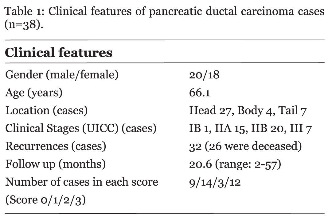
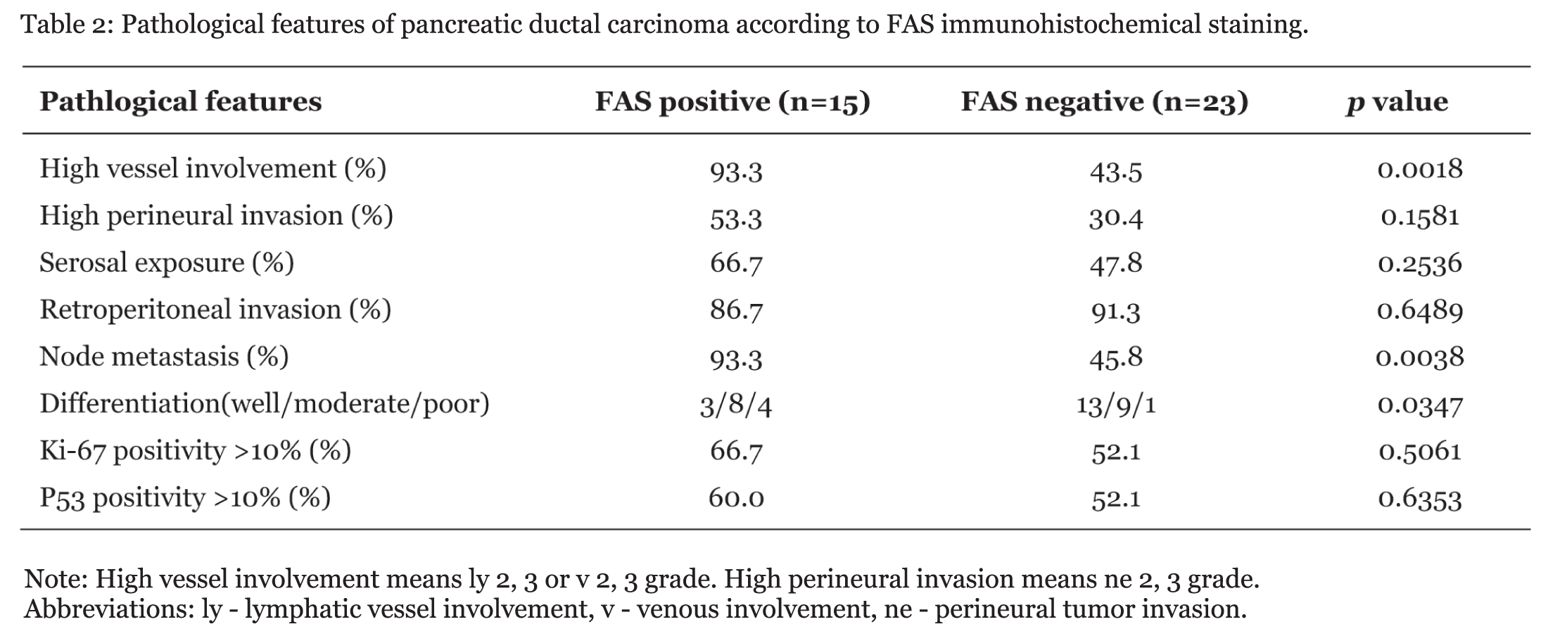
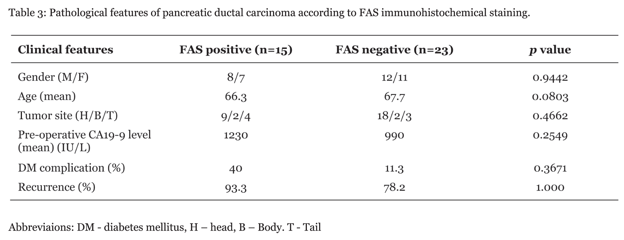
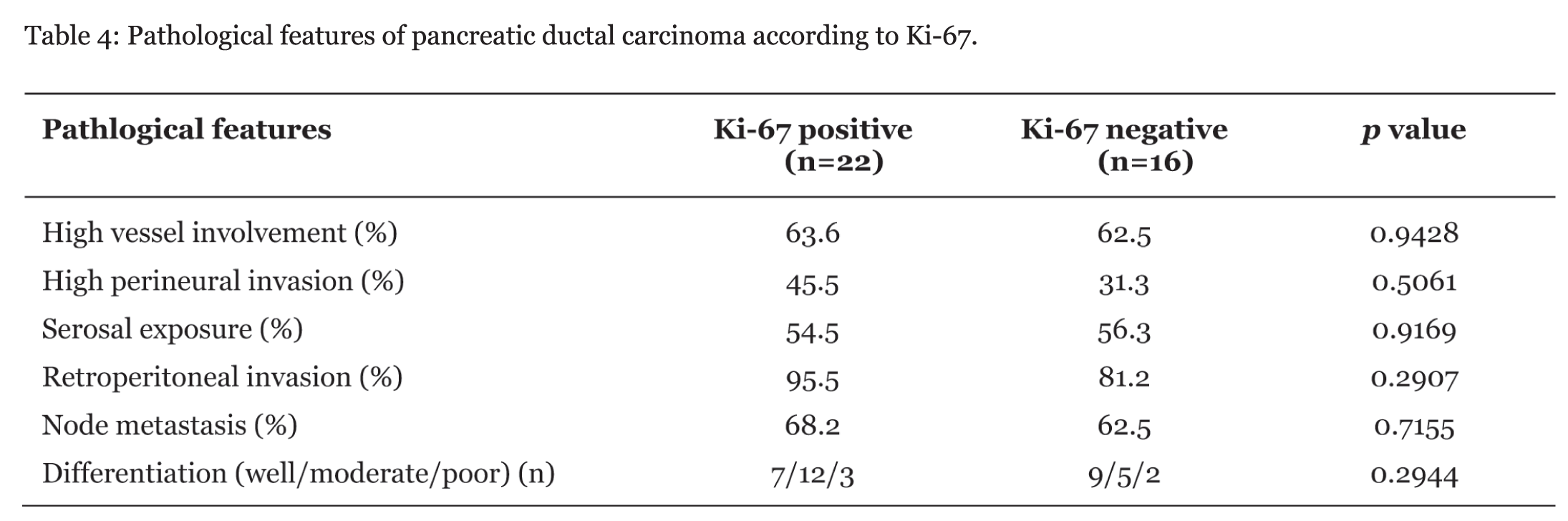
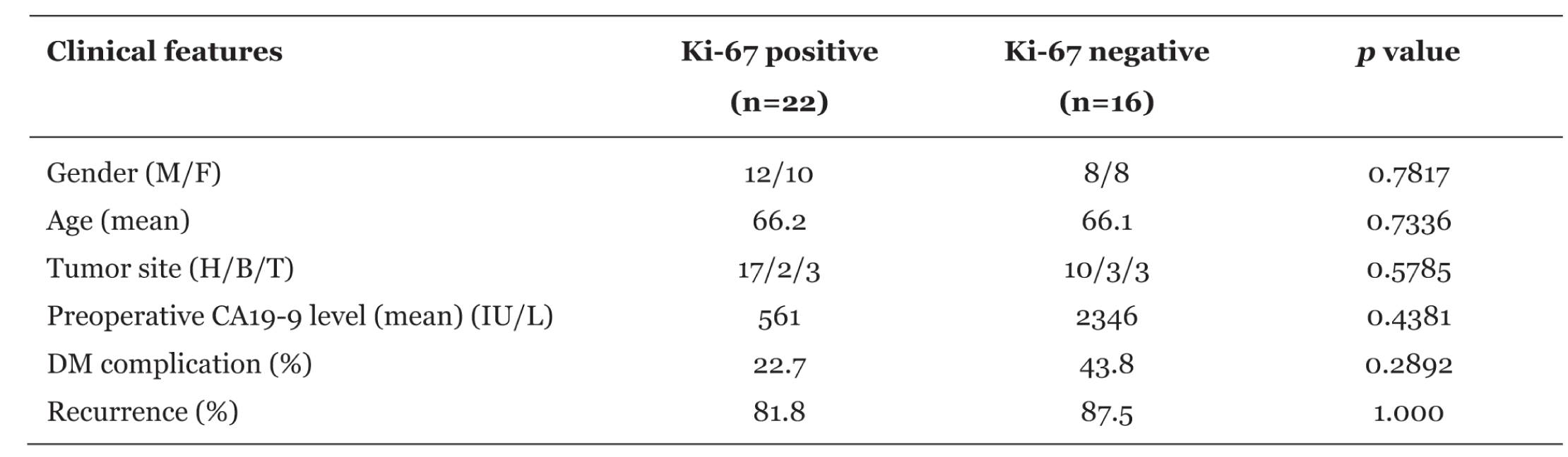
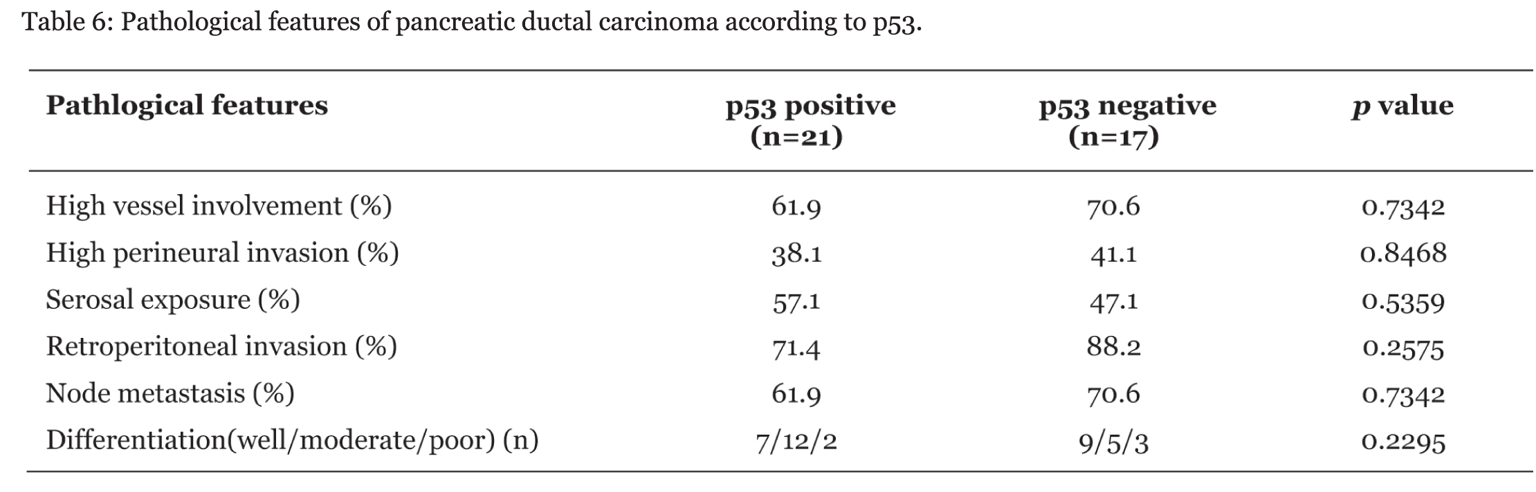
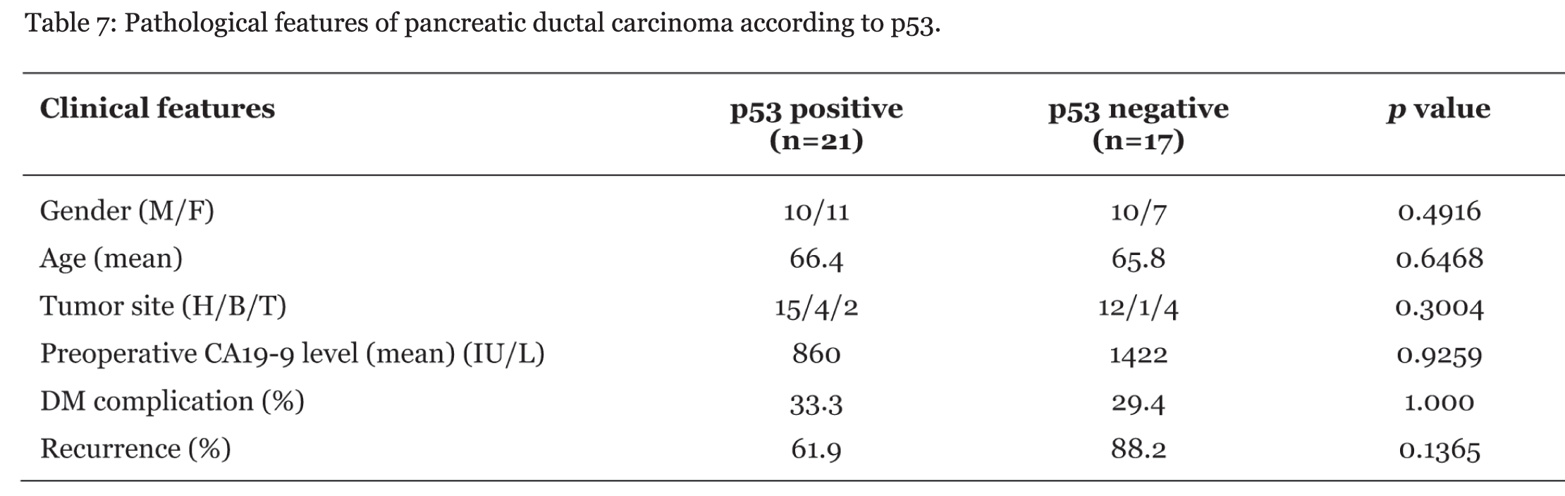
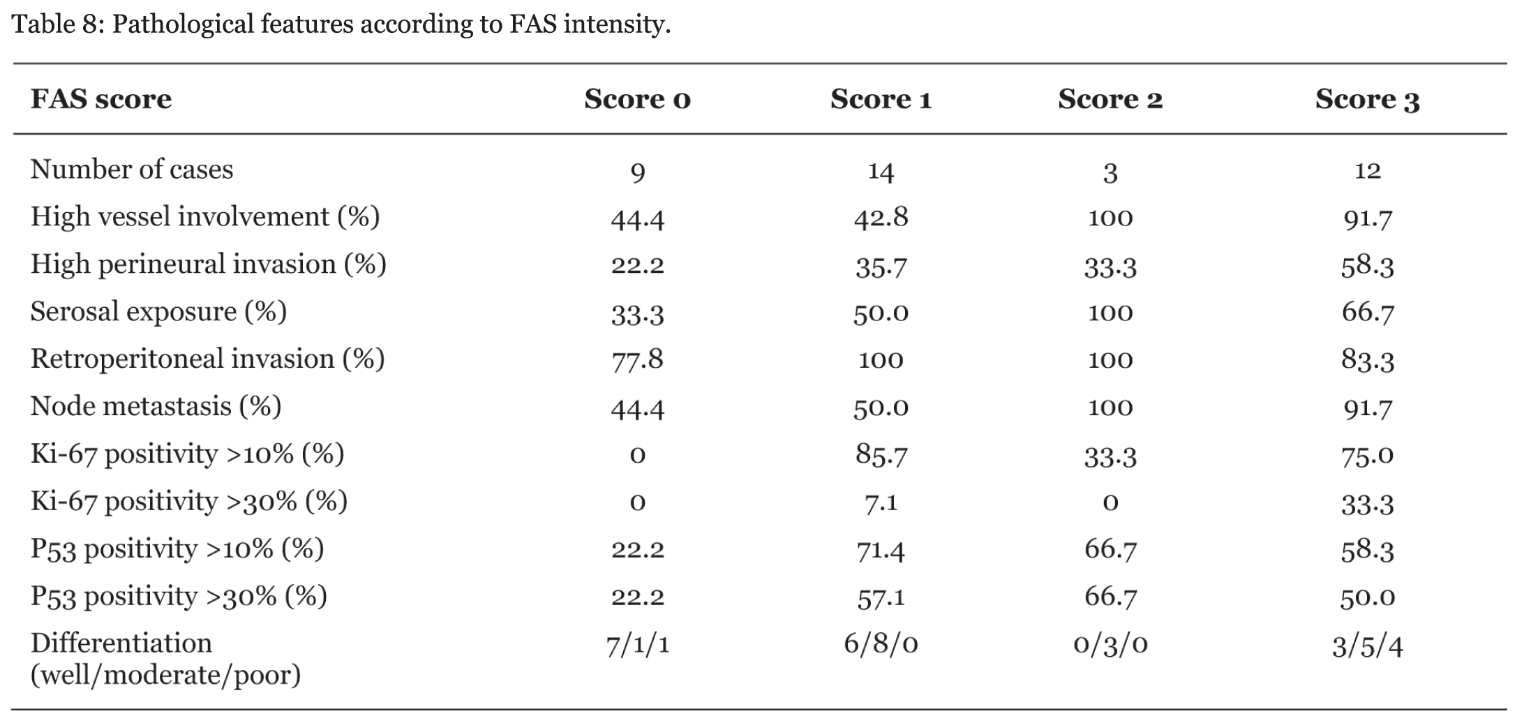
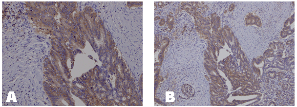
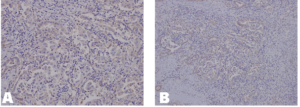
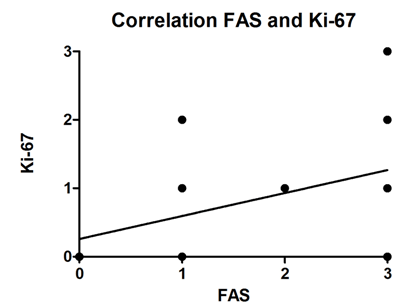
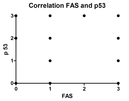
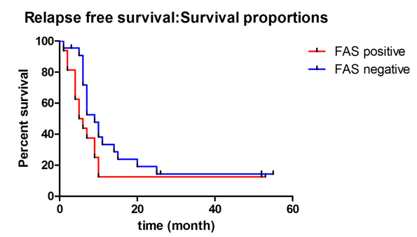
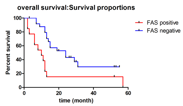
The clinical features of 38 PDC cases are listed in table 1. All 38 cases had adenocarcinoma and recurrences were noted in 32 patients. Twenty six cases died due to PDC. Fifteen cases were positive and 23 were negative for FAS staining (Figures 1, 2). Correlation between scores of tissue FAS expression and scores of tissue Ki-67 expression was analyzed. FAS positivity correlated with Ki-67 positivity (p = 0.001) (Figure 3). There was no relationship between scores of tissue FAS expression and scores of tissue p53 expression (p = 0.2240) (Figure 4). The pathological features were analyzed according to FAS expression status. FAS positive cases showed higher rates of high vessel involvement (p = 0.001), lymph node metastases (p = 0.004) and differentiation (p = 0.035) (Table 2). The clinical features of the two groups were also analyzed. There were no significant differences between the two groups in gender, age, tumor site, preoperative levels of CA19-9 or diabetes mellitus complication (Table 3). Clinicopathological findings are shown according to Ki-67 or p53 positivity (Tables 4, 5). There were no differences in the clinicopathological features according to positivity of Ki-67 or p53. Although recurrences were often detected in both groups, there was no difference in relapse free survival rates (RFS) between the two groups (p = 0.1499) (Figure 5). However, overall survival rate (OS) of FAS positive cases was lower than that of FAS negative cases (p = 0.007) (Figure 6) | ||||||
| ||||||
|
| ||||||
| ||||||
|
| ||||||
| ||||||
|
| ||||||
| ||||||
|
| ||||||
| ||||||
|
| ||||||
| ||||||
|
| ||||||
| ||||||
|
| ||||||
| ||||||
|
| ||||||
|
| ||||||
|
| ||||||
|
| ||||||
|
| ||||||
| ||||||
|
| ||||||
|
| ||||||
|
| ||||||
|
| ||||||
|
| ||||||
|
| ||||||
|
Discussion
| ||||||
|
Human FAS is a 270-kDa cytosolic enzyme that synthesizes long-chain fatty acids, palmitate, myristate, and stearate, using acetyl-CoA, malonyl-CoA, and NADPH. [9] Recently, it has been suggested that FAS plays an important role not only in tumor growth, but also in tumor survival and drug resistance with de novo lipogenesis. [18] [19] Therefore, increase in FAS expression may be associated with cancer progression, high risk of cancer recurrence, and shorter survival. Pancreatic ductal cancer is known to be a cancer with poor prognosis because recurrences are often seen even though curative resections are performed. Recently, several studies of FAS expression in cases of pancreatic ductal cancer have been reported. [17] [19] [20] In these studies, FAS was reported to have relationship to some pathological characteristics such as histological type of cancer, lymph node metastases, p53 expression or clinical outcome. Alo et al. [17] reported that FAS was overexpressed in poorly differentiated adenocarcinomas and was associated with patients' outcome in pancreatic adenocarcinoma. Witkiewicz et al. [20] reported that FAS expression strongly correlated with tumor size, tumor grade and the presence of lymph node metastasis in pancreatic cancer. Our results were similar to their conclusions, but, interestingly, FAS positivity had no corelation with p53 positivity, though FAS positivity corelated with Ki-67 positivity. Positivity for p53 immunohistochemical staining is reported to be related to clinical outcome of different types of cancers. D'Erchia et al. [21] demonstrated that p53 family of proteins control FASN expression, the same as the SREBP-1c signalling pathway. In contrast, Ogino et al. [16] reported that FAS expression was associated with clinical outcome, but it had no significant relationship with p53 positivity. Rashid et al. [22] also reported FAS staining in 139 cases of colorectal cancer, and suggested that FAS expression could correlate with poor prognosis through an association with lymph node metastases and aggressive histopathological subtypes, however, no significant association was observed between lymph node metastases, patient survival, and FAS expression. Our results of correlation between FAS expression and p53 expression was similar to Ogino's et al. From the previous reports and our results, it is proposed that FAS expression is related to cancer aggressiveness. FAS positivity may be considered to be one of the prognostic markers. However, the results of FAS expression are slightly different among previous reports. We suspect that these differences may be due to differences in criteria for positivity for FAS and p53 staining. More studies are needed to analyze FAS expression for establishing the exact singnificance of FAS expression. | ||||||
|
Conclusions
| ||||||
|
FAS expression may be related to clinical behavior in pancreatic ductal carcinoma with high incidence of lymph node metastasis. FAS positive cases of pancreatic ductal carcinoma may have shorter survival time compared to FAS negative cases. | ||||||
|
References
| ||||||
| ||||||

|
[HTML Abstract]
[PDF Full Text]
|
|
Author Contributions:
Hiroshi Maekawa - Conception and design, Acquisition of data, Analysis and interpretation of data, Drafting the article, Critical revision of the article, Final approval of the version to be published Tomoaki Ito - Conception and design, Acquisition of data, Analysis and interpretation of data, Drafting the article, Critical revision of the article, Final approval of the version to be published Hajime Orita - Conception and design, Acquisition of data, Analysis and interpretation of data, Drafting the article, Critical revision of the article, Final approval of the version to be published Koichi Sato - Conception and design, Acquisition of data, Analysis and interpretation of data, Drafting the article, Critical revision of the article, Final approval of the version to be published Ryo Wada - Conception and design, Acquisition of data, Analysis and interpretation of data, Drafting the article, Critical revision of the article, Final approval of the version to be published |
|
Guarantor of submission:
The corresponding author is the guarantor of submission. |
|
Source of support:
None |
|
Conflict of interest:
Authors declare no conflict of interest. |
|
Copyright:
© Hiroshi Maekawa et al. 2013; This article is distributed the terms of Creative Commons attribution 3.0 License which permits unrestricted use, distribution and reproduction in any means provided the original authors and original publisher are properly credited. (Please see Copyright Policy for more information.) |
|
|