| Table of Contents |  |
|
Review Article
|
| A review of transplantation for tumors other than hepatocellular carcinoma |
| Ryan Ruel T Barroso1, Daniel Cherqui2, |
|
1Department of Surgery, Cardinal Santos Medical Center, San Juan, Manila, Philippines.
2Chief - Section of Hepatobiliary Surgery & Liver Transplantation New York Presbyterian - Weill Cornell Medical College, New York, USA. |
|
Article ID: 100008IJHPDRRTB2012 doi:10.5348/ijhpd-2012-8-RA-6 |
|
Address correspondence to: Ryan Ruel T Barroso, MD Rm 215, Medical Arts Bldg, Cardinal Santos Medical Center Wilson St., San Juan Manila Philippines Phone: +63 9175540024 Email: RyeBarroso@gmail.com |
|
[HTML Abstract]
[PDF Full Text]
|
| How to cite this article: |
| Barroso RRT, Cherqui D. A review of transplantation for tumors other than hepatocellular carcinomat. International Journal of Hepathobiliary and Pancreatic Diseases 2012;2:24–30. |
|
Abstract
|
|
Introduction:
Debates are ongoing in many centers for the indications of liver transplantation for non-hepatocellular carcinoma tumors due to a number of factors. Standardized protocols are lacking and clinical outcomes vary.
Objective: A review was performed of the numerous published articles, with focus on the management and clinical outcome of tumors besides hepatocellular carcinoma (HCC) which were treated by liver transplantation, namely, cholangiocarcinoma, neuroendocrine tumors and hepatic epitheloid hemangioendothelioma in an attempt to promote a more standardized guideline. Discussion: Liver transplantation for cholangiocarcinoma reveals promising results in selected group of patients who underwent neoadjuvant chemoradiation prior to transplantation. For neuroendocrine tumors with liver metastases a number of selection criteria have been proposed in different centers, however the results of which are still variable. Transplantation is a viable option for unresectable hepatic epitheloid hemangioepithelioma with results at par with resection. Conclusion: Over time as patient selection, techniques, peri-operative care and the understanding of the different diseases improves, the results of liver transplantation for these tumors likewise will became favourable. However, standardization of the management and guidelines are still needed. | |
|
Key Words:
Liver Transplant, Cholangiocarcinoma, Neuroendocrine Tumors, Hepatic Epitheloid Hemangioepithelioma, Clinical Outcome.
| |
|
Introduction
| ||||||
|
The indication for liver transplantation in tumors beyond hepatocellular carcinoma (HCC) are still under debate because of variable results reported in available literature, lack of standardized approach, no standard criteria for patient selection and shortage of donor grafts. Historically, liver transplantation for tumors other than HCC emerged as a desperate option, but recently, as knowledge and technique expands, more and more data has emerged proving that liver transplantation is indeed a valid therapeutic modality. The authors present this review of literature with emphasis on the clinical outcomes of liver transplantation for tumors other than HCC, specifically for cholangiocarcinoma, neuroendocrine tumors and hepatic epitheloid hemangioendothelioma. Cholangiocarcinoma: Summary of results of liver transplantation for cholangiocarcinoma is shown in table 1. Cholangiocarcinoma (CCA) is an uncommon but highly aggressive group of malignant tumors arising from the biliary duct epithelium. [1] It is the second most prevalent primary cancer of the liver, with a slight male preponderance and no identifiable etiologic factor in most patients. [2] It is however, associated with chronic biliary inflammation such as in the case of primary sclerosing cholangitis. Other risk factors include: age, diabetes, stones in the biliary duct, liver cirrhosis, choledochal cysts, caroli's disease, parasitic infections of the liver and other drugs and toxic chemicals. [3] CCA comprise <3% of all malignant tumors, and have a conspicuous demographic variation with the highest incidence in South East Asia. In the US, about 3000-5000 cases are reported per year [4] with a prevalence of 1.2 per 100,000. [5] There are two major classifications of CCA according to anatomic site: Intrahepatic and Extrahepatic. [1] [6] [7] Intrahepatic CCAs, (5–10% of all cases) are usually mass forming tumors arising from the intrahepatic ducts. Extrahepatic CCAs which can arise from anywhere along the extrahepatic duct system can be further subclassified into proximal (hilar and perihilar) and distal tumors. The proximal tumors (60–70% of all cases) usually present with a sclerosing type of growth pattern while the distal tumors (20–30% of all cases) usually present as polypoid masses. [7] To date, the unavailability of screening systems hampers the diagnosis of CCA at an early stage and therefore contributes to the dismal outcome of this disease. Most of the patients with unresectable tumors succumb within 6–12 months (despite non-surgical treatment). Curative surgical resection remains the standard of treatment except for patients with primary sclerosing cholangitis wherein the presentation is usually multifocal hence the relatively high (>90%) recurrence rate. [8] The prognosis for surgical resection depends upon the anatomic location of the tumor. Five- year survival rate of intrahepatic CCA is 22–40%, hilar cholangicarcinoma is 11–41% and for distal extrahepatic tumors is 27–37%. [9] Liver transplantation offers a vivified approach since it deals with the diseased liver as in the case of PSC, as well as with unresectable disease allowing for tumor-free margins. Regardless, the historical results from transplantation afforded very little hope. Reported five years survival rate after liver transplantation was between 0–18% for intrahepatic CCA and 23–26% for extrahepatic CCA [10] [11] [12] [13] not to mention the high recurrence rates. In 2000, Meyer and collegues confirmed such poor results by reviewing the Cincinnati Transplant Tumor Registry database. Two hundred seven patients who underwent orthotopic liver transplant (OLT) for cholangiocarcinoma between 1968 to 1997 were analyzed and the five years survival rates were only 23% with recurrence rate of 51% within a year. For this reason, CCA was considered a contraindication to liver transplantation in most transplant centers. In 2002, Sudan and colleagues from the University of Nebraska published a report on radiochemotherapy prior to OLT. Their protocol, which was implemented in 1987 enrolled a total of 16 patients with hilar CCA and one patient with distal CCA in the setting of primary sclerosing cholangitis. They were given neoadjuvant radiochemotherapy and 11 of the patients (after excluding those with extrahepatic disease) underwent liver transplant. Five out of the 11 patients were alive and free from tumor recurrence 2.8–14.5 years after transplantation leading to the possibility of long term survival in highly selective cases. [14] In 2005, Rea and collegues published a retrospective report comparing neoadjuvant chemoradiation+OLT to surgical resection for patients with stage 1 and 2 hilar cholangiocarcinoma. Although it is quite a task to compare the results between the resection and the transplant cohorts, the transplant results were quite substantial. In the 38 patients who underwent OLT, they were able to achieve 90%, 82% and 82% respectively for the one-, three- and five-year survival rates. [15] In 2007, Becker and colleagues analyzed 280 patients from the UNOS database with CCA treated with liver transplant (1987–2005). The network database did lack certain details pertaining to the use of neoadjuvant therapy, nevertheless, their results for the one- and five- year survival rate were 74% and 38% respectively. They also found that concomitant primary sclerosing cholangitis did not have any statistically significant influence on the outcome. [16] Recently in 2011, Panjala and colleagues from the Mayo Clinic Florida published their experience with neoadjuvant chemo-radiation therapy given prior to OLT. The Mayo Clinic Protocol critically selected patients that are least likely to develop metastatic disease and patients that are most likely to respond to neoadjuvant treatment and have a high chance of survival after transplantation. Patients without extrahepatic metastases or gallbladder involvement underwent a staging operation prior to transplantation. A total of 22 patients were enrolled and all of them were able to finish the Mayo protocol and undergo liver transplantation from 2001–2008. The one-, two- and three-year Kaplan-Meier survival probability was 90%, 70% and 63% respectively. [17] Despite these very encouraging results, transplantation for CCA remain controversial. A number of important factors such as scarcity of donor grafts preclude the acceptance of liver transplantation for CCA in most centers. In the centers where CCA is a valid indication for transplantation, issues on allocation of deceased donor liver grafts remains a problem. | ||||||
| ||||||
|
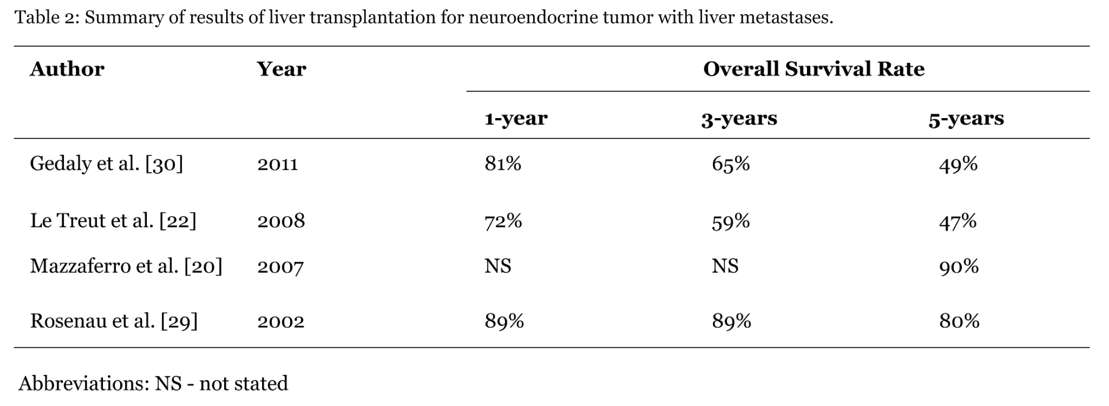
Neuroendocrine Tumors: Summary of results of liver transplantation for neuroendocrine tumors is shown in table 2. Neuroendocrine tumors (NET) comprise of a diverse group of tumors that arise from the neuroendocrine system. [18] These tumors, which were previously regarded as rare, have been reported to have a linearly increasing incidence by a number of centers (5.25 cases per 100,000) in the US (SEER 2004). [19] Perhaps these numbers are a result of improved diagnostic modalities, better screening or simply a true change of incidence, nevertheless the treatment guidelines available to date are poorly defined and the outcome, still inconsistent. NET can be classified either as a functioning tumor with symptoms caused by specific hormones being autonomously secreted or non-functioning tumors that do damage by mass effect. They can also be classified according to their occurrence, whether sporadic (gastroenteropancreatic tumors, paragangliomas, pheochromocytomas, small cell, adrenal gland tumors) or by inheritance (MEN, VHL syndrome, neurofibromatosis type 1). The most common neuroendocrine tumors are the gastroenteropancreatic tumors with a global incidence of about 5–7 cases per 100,000 [20]. The usual sites of dissemination are the liver (40–93%), bone (12–20%) and lung (8–10%). [20] [21] The presence of liver metastases are considered by many authors to be the most contemptuous prognostic factor in this disease with a five-year survival rate of approximately 13–15% if left untreated. [18] [21] Among the number of therapeutic options for NET with liver metastases (transarterial chemoembolization, radioembolization, cryoembolization, peptide receptor radiodnuclide therapy), surgical resection remains the treatment of choice. [22] But the fact that up to 80% are unresectable upon presentation coupled by the indolent nature of this disease providing a 'therapeutic window', [23] [24] reroutes us towards liver transplantation as a worthwhile option to achieve cure or palliation. The first reports on liver transplant for NET with liver metastases were published in 1988 by O'Grady et al. (two patients), and by Iwatsuki et al. (five patients). Their results were recurrence-free five-year survival of 36–57% and 24–45% respectively. However, relative lack of uniformity in patient selection made it difficult to analyze the results. [25] [26] In 1997, Le Treut et al. published a 31-case French multicentric report showing significant difference in survival between carcinoid and non-carcinoid tumors. The five-year survival of carcinoid was reported to be 69% while the four-year survival of non-carcinoid tumors was 8%. [27] Lehnert T, in 1998 published an analysis of 103 patients. Two risk factors were identified for poor survival in NET: i) extended procedures, and ii) age >50 years. Results showed 65% five-year survival without the two factors, 32% with at least one factor and 0% when both factors were present. [28] Rosenau et al. in 2002 published a report whose aim was to identify predictors of long-term survival based on tumor biology. They showed that both Ki 67 which is a nuclear protein strictly associated with cell proliferation and E-cadherin which is important in maintenance of normal tissue architecture, have significant prognostic value. They found that Ki 67% >5% and/or aberrant E-cadherin resulted in 0% 7-year survival whereas <5% and regular E-cadherin expression demonstrated 100% seven years survival. [29] Notably, the most promising results were reported by Mazzaferro et al. in 2007. In a series of 24 patients, they achieved a 77% five-year disease-free survival rate and a 90% five-year overall survival rate. They developed the Milan criteria for indication of liver transplantation in patients with hepatic metastases and their selection criteria included the following: i) patients =55 years of age with low grade tumors, ii) primary tumor being drained by the portal system, iii) removal of the primary tumor must be done by a curative resection through a separate operation from the OLT, iv) metastatic diffusion to liver parenchyma is 50% or less, and v) the disease must be stable for at least six months during the pre-transplantation period. [20] About a decade after their first publication on the topic Le Treut et al. published an 85-case multicenter report in 2008 and they were able to identify other predictors of long term survival. Their study showed a substantial difference in overall survival, favoring patients with non-duodenopancreatic tumors and limited hepatic disease (68% five years survival) over patients with hepatomegaly and primary duodenopancreatic tumors (12%). [22] In 2011, Gedaly and collegues published a retrospective analysis of 150 patients from the UNOS database who underwent LT for NET with liver metastases. They noted that the patient's age, which was previously a factor in a number of selection criterias was found to have no statistical significance in the treatment outcome and that the one-, three-, and five-year survival rate of NET with metastases had similar outcome with patients with HCC metastasis who underwent OLT. Interestingly their analysis pointed out that patients waiting for more than two months on the listing had significantly better outcome. [30] To date there are no published randomized controlled trials comparing the different therapeutic modalities for the treatment of NET with liver metastases. Liver transplantation remains a promising option for patients with disseminated disease confined to the liver. A multidisciplinary approach to the management of NET is of utmost importance as well as the need to define a more precise criterion for patient selection. | ||||||
| ||||||
|
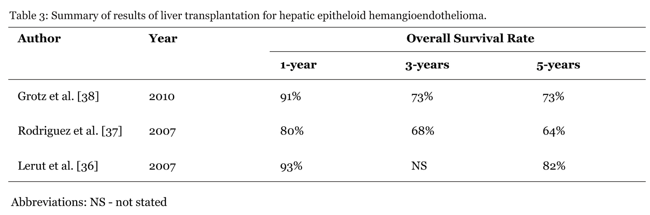
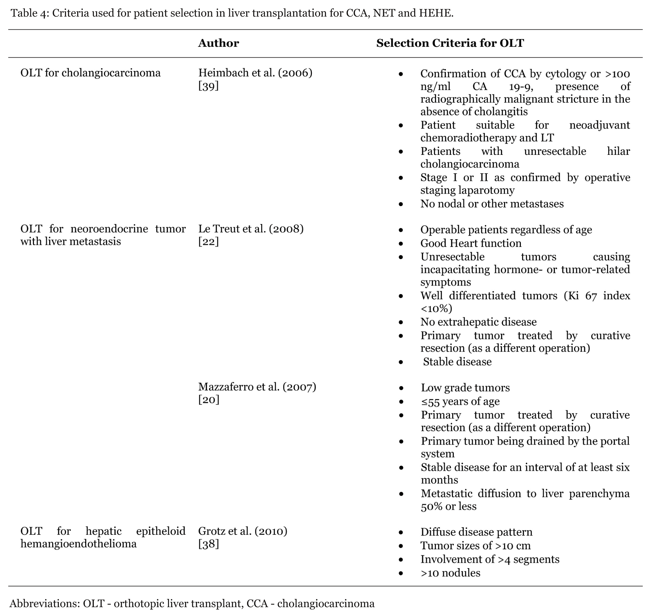
Hepatic Epithelioid Hemangioendothelioma: Summary of results of liver transplantation for hepatic epitheloid hemangioendothelioma is shown in table 3. Hepatic epitheloid hemangioendothelioma (HEHE) is the more common form of epitheloid hemangioendothelioma, a rare, low grade, soft tissue tumor that arises from the vascular endothelium which can involve the liver, lungs, spleen, stomach and heart. It has a 3:2 female preponderance with a peak incidence between the ages of 30–40 years. [31] There are no clearly defined risk factors for developing HEHE although some studies report the following factors may contribute to its development: primary biliary cirrhosis, alcohol abuse, liver trauma, viral hepatitis, oral contraceptives, vinyl chloride and asbestos. It has a variable malignant potential with an unpredictable clinical course and the reported incidence is one per million population. [32] In a study of 402 patients done by Mehrabi et al. in 2006, on diagnosis, patients most frequently presented with right upper quadrant pain (48.6%), weight loss (15.6%) and an enlarged liver on physical examination (20.4%). Other signs and symptoms included weakness, ascites, emesis, jaundice and fatigue. About 25% patients were asymptomatic. In the same study they noted that about 87% patients had multifocal, bilobar tumors and only 13% patients presented with unifocal disease. They observed that in either presentation there was tendency towards involvement of the right lobe. Extrahepatic involvement was observed in only 36.6% patients at the time of diagnosis. [33] Imaging studies for HEHE shows that the lesions are more similar to metastatic carcinoma rather than vascular lesions. [34] When diagnosed early they may present as multiple nodules of about 0.5–12 cm in diameter whereas at a later stage these nodules tend to coalesce and form a more diffuse type of lesion commonly in the periphery of the liver. [35] In 2007, the European Liver Transplant Registry (ELTR) reported 1, 5 and 10-year survival rates of 93%, 82% and 72% with diseases free survival of 90%, 82% and 64% respectively (n=59 patients). It is important to note their observation that lymphnode status, neoadjuvant therapy or the presence of extrahepatic disease did not have any statistical influence on the overall survival. Microvascular involvement on the other hand was found to have a significant negative influence over the disease free survival. [36] In the same year, Rodriguez et al. reviewed the UNOS database of 110 patients who underwent liver transplant between 1987 to 2005. Their one-, three- and five-year overall survival rates were 80%, 68% and 64% respectively. [37] The Mayo clinic, in 2010, reviewed their experience with 30 patients to compare the overall survival and disease free survival between OLT and liver resection cohorts. Liver resection was found to be associated with a one-, thre-, and five-year overall survival of 100%, 86% and 86% and a disease free survival rate of 78%, 62% and 62% respectively. For the group that underwent OLT, they were associated with an overall one-, three-, and five-year survival rates of 91%, 73% and 73% and disease free survival rate of 64%, 46% and 46%. They were able to identify certain clinicopathological factors with prognostic values such as tumor size and multifocal disease. Tumor sizes of =10 cm as well as involvement of =4 liver segments were found to have better disease free survival (p=0.003 and p=0.02 respectively) compared to larger tumors and tumors involving a larger part of the liver. Tumor nodules =10 in number also appears to have positive effect on the disease free survival (p=0.052). [38] A management algorithm was designed by their group: Those with the previously mentioned factors should undergo liver resection whereas those patients with more diffused disease pattern, tumors >10 cm in size, involvement of >4 segments and >10 tumor nodules should undergo OLT. Because of the rarity of this disease there are neither specific guidelines nor a standardized therapeutic approach to its management. The results of OLT for HEHEs are favorable and should therefore be considered as a first line therapy for unresectable disease. The therapeutic strategies should be tailored specifically for each patient and their unique presentation. The criteria used for patient selection in liver transplantation for cholangiocarcinoma, neuroendocrine tumors and hepatic epithelioid hemangioendothelioma are shown in table 4. | ||||||
| ||||||
|
| ||||||
| ||||||
|
Conclusion
| ||||||
|
Liver transplantation for the treatment of non-HCC tumors shows encouraging results especially over time as we understand more about each individual disease entity, and improve on the technical aspects as well as pre- and post-surgical care. Early diagnosis, meticulous patient selection, and a multi-disciplinary approach towards the overall management of these diseases are key factors for a good outcome. | ||||||
|
References
| ||||||
| ||||||


|
[HTML Abstract]
[PDF Full Text]
|
|
Author Contributions:
Ryan Ruel Tupas Barroso - Conception and design, Acquisition of data, Analysis and interpretation of data, Drafting the article, Critical revision of the article, Final approval of the version to be published Daniel Cherqui - Conception and design, Acquisition of data, Analysis and interpretation of data, Drafting the article, Critical revision of the article, Final approval of the version to be published |
|
Guarantor of submission:
The corresponding author is the guarantor of submission. |
|
Source of support:
None |
|
Conflict of interest:
Authors declare no conflict of interest. |
|
Copyright:
© Ryan Ruel Tupas Barroso et al. 2012 This article is distributed the terms of Creative Commons Attribution License which permits unrestricted use, distribution and reproduction in any means provided the original authors and original publisher are properly credited. (Please see Copyright Policy for more information.) |
|
|