|
Case Report
Unveiling the enigma of spontaneous regression in hepatocellular carcinoma: A case report from a tertiary care center in South India
1 Ida B. Scudder Cancer Centre, Department of Radiation Oncology, Christian Medical College, Vellore, Tamil Nadu, India
2 Department of Radiodiagnosis, Christian Medical College, Vellore, Tamil Nadu, India
3 Department of Hepatobiliary and Pancreatic Surgery, Christian Medical College, Vellore, Tamil Nadu, India
4 Department of Pathology, Christian Medical College, Vellore, Tamil Nadu, India
5 Department of Hepatology, Christian Medical College, Vellore, Tamil Nadu, India
Address correspondence to:
Thomas Samuel Ram
Ida B. Scudder Cancer Centre, Department of Radiation Oncology Unit 1, Christian Medical College, Vellore, Tamil Nadu,
India
Message to Corresponding Author
Article ID: 100105Z04RK2024
Access full text article on other devices

Access PDF of article on other devices

How to cite this article
Benny RK, Tony V, Thanikaivelu S, Raju RS, Rijo Isaac NP, Barman B, Kiruba A, Thomas RS, John NO, Sathyamurthy A, Ramireddy JK, Ram TS. Unveiling the enigma of spontaneous regression in hepatocellular carcinoma: A case report from a tertiary care center in South India. Int J Hepatobiliary Pancreat Dis 2024;14(2):15–26.ABSTRACT
Introduction: Spontaneous regression in hepatocellular carcinoma (HCC) is an enigma as it is a very rare phenomenon, multiple possible hypotheses were described to support this enigma.
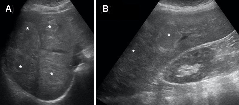
Case Report: A 61-year-old man known Type 2 diabetes mellitus and hypertension was evaluated for complaints of unexplained weight loss (40 kg loss in eight months), loss of appetite, along with generalized weakness of three months duration. He underwent computed tomography (CT) scan abdomen that showed arterial phase hyper-enhancing lesion (white short arrow) in left lobe/segment V of liver with washout. His alpha-fetoprotein (AFP) was 12263 IU. He was advised transarterial radioembolization (TARE) and systemic therapy. He did not undergo any treatment due to logistical issues. After three months he underwent a repeat CT scan, which showed decrease in the size of the heterogeneously hypodense space-occupying lesion (SOL) with wall irregularity involving liver segments II, III, IV, and V. His AFP level had fallen to 600 IU. He underwent a diagnostic laparoscopy, intraoperative ultrasound scan, frozen section (a rapid intraoperative histopathological diagnosis) proceeds left hepatectomy (including distal middle hepatic vein) and excision of 2 lesions in the caudate lobe and cholecystectomy under general anesthesia. The left hepatectomy specimen showed a scanty viable tumor (~5%) consistent with moderately differentiated hepatocellular carcinoma and with secondary changes (~95%), including extensive necrosis, xanthogranulomatous inflammation, and hemorrhage. He was followed up for three years with serial CT scan and was found to be disease free with 3 years AFP value of 1.32 IU.
Conclusion: We conclude that partial spontaneous resolution of hepatocellular carcinoma (HCC) is rare but a possible phenomenon with multiple mechanisms explaining the enigma and it presents an opportunity for further research. The collection and thorough analysis of clinical data obtained from patients who have experienced spontaneous resolution of HCC will help understand this mysterious phenomenon. It could also lead to the development of new treatment strategies for HCC based on the possible hypothesis.
Keywords: AFP, Biopsy proven, Hepatocellular carcinoma, Spontaneous resolution
SUPPORTING INFORMATION
Acknowledgments
RBK would like to thank SGG for proof reading, AKK and ABK for all the help.
Author ContributionsRajendra Benny K - Substantial contributions to conception and design, Acquisition of data, Analysis of data, Interpretation of data, Drafting the article, Revising it critically for important intellectual content, Final approval of the version to be published
Vinitha Tony - Acquisition of data, Interpretation of data, Drafting the article, Revising it critically for important intellectual content, Final approval of the version to be published
Sonia Thanikaivelu - Drafting the article, Revising it critically for important intellectual content, Final approval of the version to be published
Ravish Sanghi Raju - Interpretation of data, Revising it critically for important intellectual content, Final approval of the version to be published
Rijo Isaac NP - Drafting the article, Revising it critically for important intellectual content, Final approval of the version to be published
Bedanta Barman - Interpretation of data, Drafting the article, Revising it critically for important intellectual content, Final approval of the version to be published
Allen Kiruba - Drafting the article, Final approval of the version to be published
Rohan Samuel Thomas - Interpretation of data, Revising it critically for important intellectual content, Final approval of the version to be published
Neenu Oliver John - Interpretation of data, Revising it critically for important intellectual content, Final approval of the version to be published
Arvind Sathyamurthy - Interpretation of data, Revising it critically for important intellectual content, Final approval of the version to be published
Jeba Karunya Ramireddy - Interpretation of data, Revising it critically for important intellectual content, Final approval of the version to be published
Thomas Samuel Ram - Interpretation of data, Revising it critically for important intellectual content, Final approval of the version to be published
Guarantor of SubmissionThe corresponding author is the guarantor of submission.
Source of SupportNone
Consent StatementWritten informed consent was obtained from the patient for publication of this article.
Data AvailabilityAll relevant data are within the paper and its Supporting Information files.
Conflict of InterestAuthors declare no conflict of interest.
Copyright© 2024 Rajendra Benny K et al. This article is distributed under the terms of Creative Commons Attribution License which permits unrestricted use, distribution and reproduction in any medium provided the original author(s) and original publisher are properly credited. Please see the copyright policy on the journal website for more information.